Peer-Reviewed Preclinical Science · 20+ Years Large-Animal Expertise
Understanding the Crucial Role of a Heart Valve Replacement Animal Model
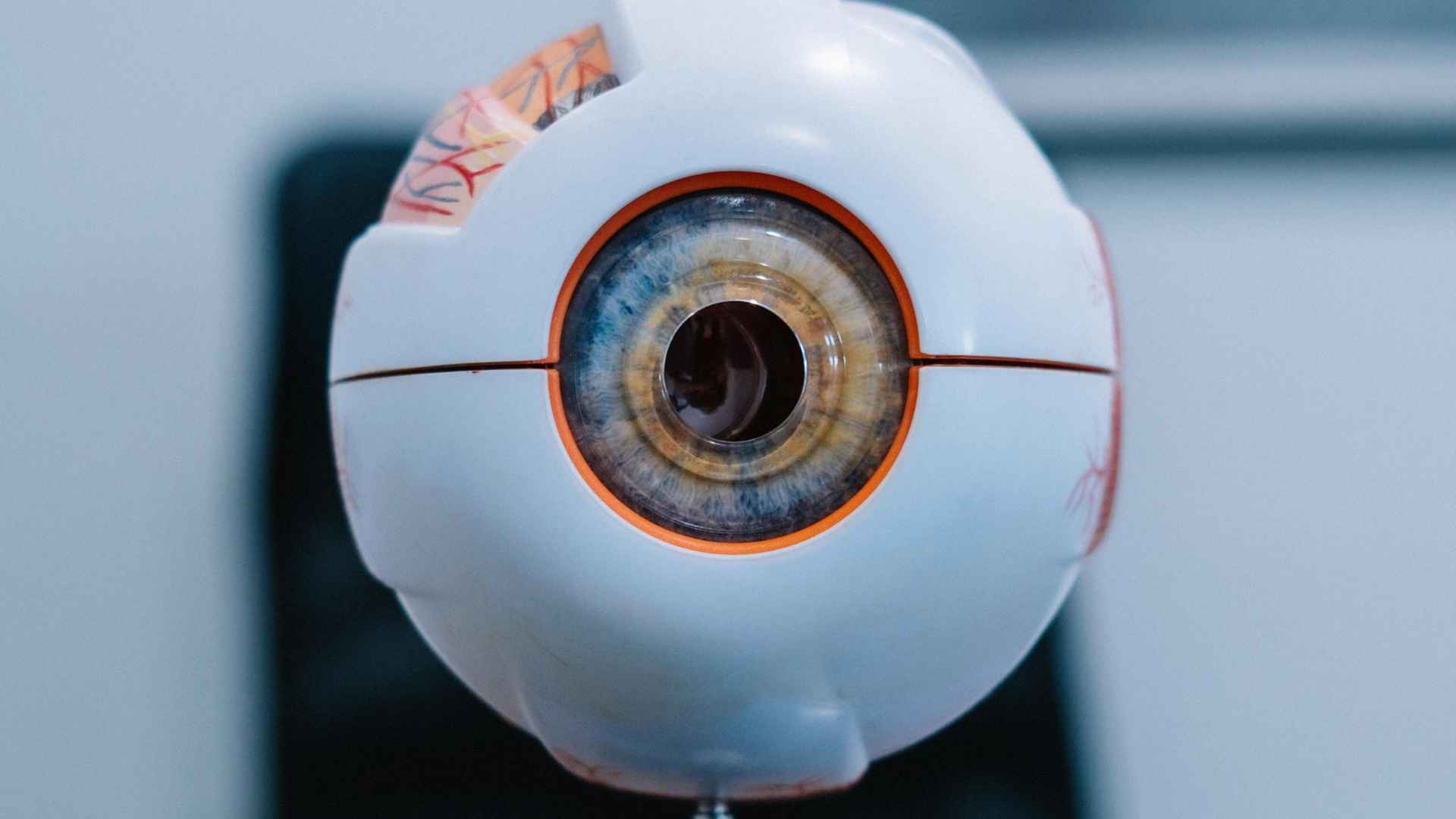
A heart valve replacement animal model is an experimental in-vivo system — typically built on large mammals such as sheep, pigs, or calves — used to evaluate prosthetic valves and transcatheter devices under physiological conditions that meaningfully resemble those of the human cardiovascular system. The purpose of the model is to gather safety, functionality, and feasibility evidence before a device enters first-in-human trials, while exposing the implant to realistic flow, pressure, and tissue responses.
Within a properly designed heart valve replacement animal model, researchers assess hemodynamics, paravalvular leakage, transvalvular gradients, leaflet kinematics, long-term durability, and biocompatibility. The same platform makes it possible to investigate phenomena that no bench loop can fully reproduce — such as thrombosis on metallic struts, pannus formation, and structural calcification of biological tissues over months of follow-up.
🔮 Expert Insight
Large-animal cardiovascular models remain the gold standard for valve device validation precisely because no computational or bench-top alternative can replicate the integrated biologic response — calcification, neointima formation, thrombus dynamics — that determines real-world device longevity. Choosing the wrong species, wrong sizing protocol, or wrong follow-up window can invalidate months of expensive work. Precision at the protocol stage pays dividends at every subsequent regulatory checkpoint.
Why Large-Animal Models Became the Industry Standard for Prosthetic Valve Testing
Cardiac dimensions, stroke volume, coronary flow patterns, and aortic root geometry in sheep and pigs approximate those of adult humans far better than rodent or rabbit hearts. This anatomical and hemodynamic proximity is what makes a prosthetic heart valve animal study in a sheep or pig the realistic predictor of clinical performance that regulators expect to see in a submission file.
Large animals also tolerate the full spectrum of clinical-grade procedures: median sternotomy, cardiopulmonary bypass, transapical access, and percutaneous femoral delivery. Investigators can apply transesophageal echocardiography, fluoroscopy, angiography, and CT just as in a human cath lab, as discussed extensively in the literature on Large Animal Models for Transcatheter Heart Valve Prosthesis evaluation. This translational fidelity is the reason large animal work remains a regulatory expectation even as in-silico and ex-vivo tools continue to advance.
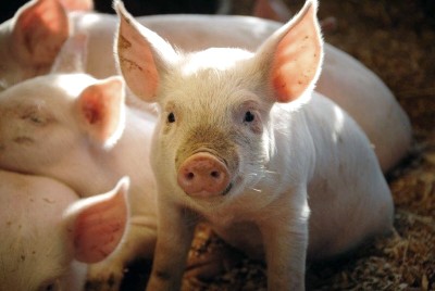
Sheep vs. Pig: Which Animal Model Is Better for Heart Valve Replacement Studies?
There is no universal winner. The selection between an ovine and a valve replacement pig model depends on the target valve position (aortic, mitral, pulmonary, tricuspid), whether the device is surgical or transcatheter, the planned study duration, and the specific failure mode under investigation. Sheep tend to calcify bioprosthetic tissue more aggressively — useful when calcification resistance is the primary endpoint — while pigs may be more relevant when thrombogenicity and rapid neointimal coverage are central concerns in TAVR preclinical testing.
🐑 Ovine (Sheep) Model
Preferred when long-term durability and biocompatibility are the central endpoints. Sheep offer heart rates, systemic pressures, and aortic annulus diameters well aligned with adult human ranges.
- Accelerated calcification biology stresses tissue coatings within months
- Stable anatomical size ideal for multi-month follow-up
- Femoral access accommodates human-sized TAVR delivery systems
- Preferred platform for TAVR preclinical testing and tissue valve biocompatibility
🍕 Porcine (Pig) Model
Preferred when acute deliverability, thrombogenicity, or precise size-matching via CT are the primary study drivers.
- CT-mapped anatomy allows very low size variability between animals
- Coagulation cascade exploitable for thrombus-focused questions
- Supports TMVR and transseptal access route development
- Challenging for long-term studies due to rapid growth altering anatomy
Comparative Snapshot: Ovine vs. Porcine Platforms
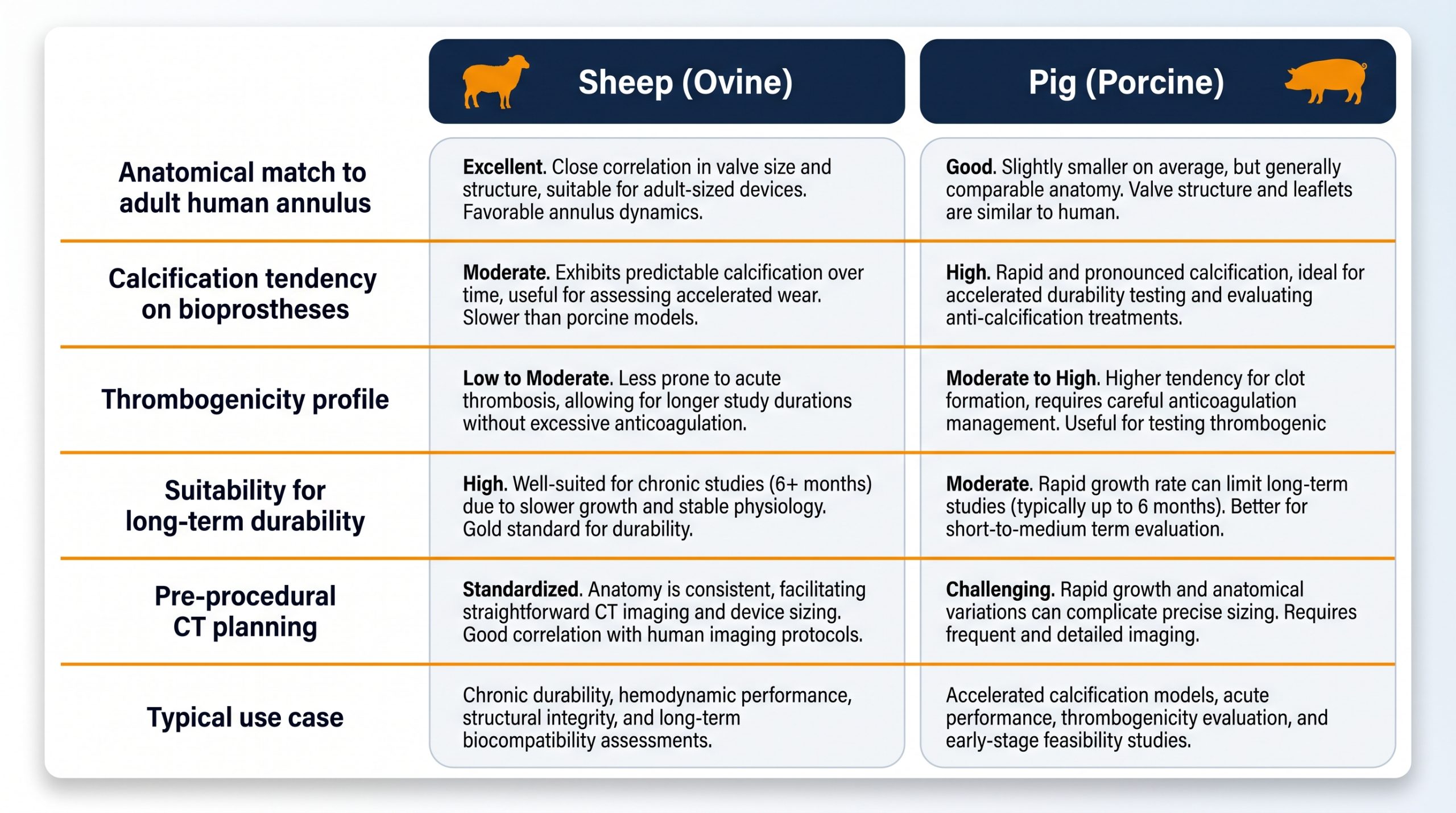
| Parameter | Sheep (Ovine) | Pig (Porcine) |
|---|---|---|
| Anatomical match to adult human annulus | Very good, stable size | Good, but rapid growth |
| Calcification tendency on bioprostheses | High (accelerated) | Moderate |
| Thrombogenicity profile | Lower spontaneous | Higher, useful for thrombus studies |
| Suitability for long-term durability | Excellent | Challenging due to growth |
| Pre-procedural CT planning | Feasible | Highly detailed and standardized |
| Typical use case | Tissue valve biocompatibility, TAVR | Acute deliverability, TMVR, thrombosis |
Practical Implementation of a Valve Replacement Pig Model
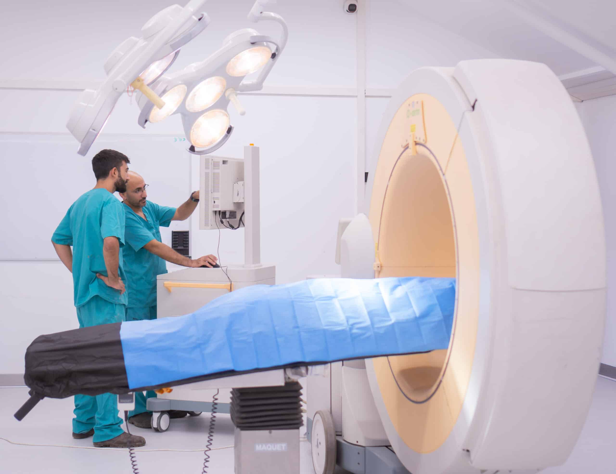
Practically, a valve replacement pig model begins with animal screening by echocardiography and contrast-enhanced CT to confirm anatomical suitability. The valve system is then delivered either surgically under cardiopulmonary bypass, or via transcatheter access through the femoral artery, apex, or transseptal route. Intra-procedural imaging confirms positioning, expansion, and absence of paravalvular leak before the animal is recovered.
Follow-up combines serial echocardiography, blood work, and ECG monitoring with a defined terminal time-point. At termination, gross pathology and histology document tissue ingrowth, leaflet integrity, and any thrombus or calcification. The same workflow supports a wide spectrum of devices — from balloon-expandable transcatheter aortic valves to dedicated mitral and tricuspid systems undergoing early transcatheter valve testing.
🔍 Protocol Note
Pre-procedural CT sizing must be performed no more than 2–4 weeks before implantation in juvenile pigs to account for growth-related annular change. Failure to re-screen can result in device-annulus mismatch that confounds hemodynamic endpoints independent of the implant’s true performance.
The Goals of TAVR Preclinical Testing: What Must the Animal Model Demonstrate?
Robust TAVR preclinical testing must answer four interlocking questions in vivo. First, deliverability and deployment: can the delivery system reach the annulus and release the valve reproducibly without injuring access vessels or the left ventricular outflow tract? Second, hemodynamic performance: are gradients low, is paravalvular leakage absent or trivial, and does cardiac output recover within the expected window?
Third, device stability: does the frame remain anchored without migration, embolization, or interference with coronary ostia? Fourth, safety: are there no unacceptable events such as conduction block, aortic injury, embolic stroke, or device thrombosis? When durability endpoints are added, the model also serves as the proving ground for novel materials — advanced by groups such as Biotech Farm, whose Israel-based facility supports tailored study designs aligned with regulatory pathways.
Deliverability & Deployment
Reproducible valve release without access-vessel injury or LVOT trauma.
Hemodynamic Performance
Low gradients, absent/trivial PVL, recovered cardiac output.
Device Stability
No frame migration, embolization, or coronary ostia interference.
Safety Profile
No conduction block, aortic injury, embolic stroke, or device thrombosis.
Key Endpoints for a Comprehensive Prosthetic Heart Valve Animal Study
A complete prosthetic heart valve animal study integrates hemodynamic, device-related, adverse-event, and pathology endpoints. Hemodynamic assessment quantifies mean and peak gradients, regurgitant fraction, effective orifice area, cardiac output, and left ventricular function. Device-related observations document position, stability, frame geometry, and structural integrity through repeated imaging.
Safety endpoints capture thrombus formation on the valve or in systemic circulation, infection, embolism, and vascular injury — endpoints that have shaped trial designs evaluating the Feasibility of Transcatheter Aortic Valve Replacement in expanding patient populations. At termination, ex-vivo histology assesses tissue ingrowth, calcification, inflammatory response, and degradation, completing the evidence package regulators expect to review.
| Endpoint Category | Key Measurements | Imaging / Method |
|---|---|---|
| Hemodynamic | Mean/peak gradient, regurgitant fraction, EOA, cardiac output | Echo (TTE/TEE), Doppler |
| Device Integrity | Frame position, expansion, leaflet motion, migration | Fluoroscopy, CT, Echo |
| Safety / Adverse Events | Thrombosis, embolism, conduction block, vascular injury | Clinical monitoring, ECG, blood work |
| Terminal Pathology | Tissue ingrowth, calcification, inflammation, leaflet wear | Gross pathology, histology, SEM |
Common Mistakes That Undermine a Heart Valve Animal Study

Several recurring errors weaken otherwise expensive studies. Choosing the wrong species for the question — for example, using growing juvenile pigs for a six-month durability study — produces artifacts that obscure real device behavior. Under-sizing or over-sizing the device relative to the animal’s annulus generates paravalvular leaks or conduction issues unrelated to the implant itself.
Other frequent issues include inadequate antithrombotic protocols, inconsistent imaging time-points, and pooling animals with very different baseline anatomies into a single dataset. A disciplined protocol, rigorous CT screening, and pre-defined exclusion criteria substantially raise the signal-to-noise ratio of any prosthetic heart valve animal study.
⚠️ Most Common Study-Invalidating Errors
- Using juvenile pigs for long-term durability studies (growth distorts annular dimensions)
- Inadequate or inconsistent antithrombotic regimens creating confounding thrombus events
- Inconsistent imaging time-points preventing meaningful longitudinal comparisons
- Pooling anatomically dissimilar animals into a single dataset without pre-defined exclusion criteria
- Skipping pre-procedural CT sizing leading to device-annulus mismatch artifacts
Duration and Follow-up for Preclinical Heart Valve Animal Studies
Study length depends on what the device must prove. Acute studies of a few days are appropriate for feasibility and deliverability. Short-term studies of one to four weeks examine early healing, initial endothelialization, and short-term function during transcatheter valve testing. Mid-term studies of one to three months track progressive tissue response and stable hemodynamics, while long-term studies of three to six months or more become essential when durability, calcification, pannus, and leaflet wear must be characterized in detail.
Regulatory expectations — including FDA guidance and ISO 5840-series standards — often dictate minimum follow-up windows for first-in-human readiness. Sustained TAVR preclinical testing over multiple months remains the principal way to generate the durability evidence those standards demand.
| Phase | Duration | Primary Questions Answered |
|---|---|---|
| Acute | 1–7 days | Deliverability, deployment, immediate hemodynamics |
| Short-Term | 1–4 weeks | Early healing, initial endothelialization, short-term function |
| Mid-Term | 1–3 months | Progressive tissue response, stable hemodynamics, pannus onset |
| Long-Term | 3–6+ months | Durability, calcification, leaflet wear, ISO 5840 readiness |
Advanced Imaging Techniques in Transcatheter Valve Preclinical Studies
Imaging is the connective tissue of modern transcatheter valve testing. Transthoracic and transesophageal echocardiography provide real-time information on leaflet motion, gradients, and regurgitation — both intra-procedurally and during follow-up. Fluoroscopy and angiography document device deployment, frame geometry, and any paravalvular jets at the moment of implantation.
Computed tomography contributes detailed 3D anatomical assessment for pre-procedural sizing and post-implantation evaluation of expansion and apposition within a valve replacement pig model. Intravascular ultrasound adds high-resolution views of the vessel wall and device-tissue interaction, supporting contrast-sparing protocols that further refine transcatheter valve testing workflows.
📺
TTE / TEE Echocardiography
Real-time leaflet motion, gradient quantification, and regurgitation assessment at all time-points including intra-procedural.
🎉
Fluoroscopy & Angiography
Device deployment documentation, frame geometry verification, and paravalvular jet detection at implantation.
📸
CT Imaging
3D pre-procedural sizing and post-implant expansion/apposition assessment — critical for porcine model studies.
🔭
Intravascular Ultrasound (IVUS)
High-resolution vessel wall and device-tissue interaction imaging; supports contrast-sparing protocols.
Addressing Complications and Ethical Considerations in Large Animal Valve Studies
Large-animal valve work carries inherent risks: anesthesia-related events, hemorrhage at access sites, myocardial injury, arrhythmias, infection, and device-related thrombosis. Mitigation depends on experienced surgical and anesthesia teams, standardized antithrombotic regimens, sterile technique, and protocolized post-operative care — including imaging that documents valve durability over time, as illustrated in clinical materials on Bioprosthetic heart valves and their failure modes.
Ethically, all such work is governed by the 3Rs — Replacement, Reduction, Refinement — and by institutional oversight. In Israel, this oversight role is carried out by the National Council for Animal Experimentation under the Ministry of Health, operating under the Animal Welfare (Experiments on Animals) Law. Facilities such as Biotech Farm align study design with these requirements from the protocol-writing stage, including documentation, severity classification, and humane endpoints.
✅ 3Rs Framework in Practice
- Replace: Use ex-vivo and computational tools at every feasible preliminary stage
- Reduce: Power calculations and strict inclusion criteria minimise animal numbers per study
- Refine: Multi-modal analgesia, humane endpoints, and optimised anaesthesia protocols reduce distress
“Every animal experiment carried out in our facility is designed with the outcome in mind: generating data that accelerates safe therapies for human patients. Animal welfare is not a checkbox — it is a scientific imperative. Healthy, unstressed animals produce more reproducible, higher-quality data.”
— Adir Koreh, CEO, Biotech Farm Ltd. & Biotech Anatomy Ltd.
Specialized Models: Creating Pathological Conditions for TAVI Evaluation
Healthy animals do not have aortic stenosis or ischemic mitral regurgitation — yet those are precisely the conditions modern devices are designed to treat. To bridge this gap, researchers create disease models. Aortic stenosis can be induced by percutaneous banding or by localised calcification techniques, producing a diseased annulus where TAVR preclinical testing better reflects the real procedural environment of high-gradient, calcified human anatomy.
For mitral interventions, controlled myocardial infarction in sheep generates functional ischemic regurgitation that mimics the clinical scenario for transcatheter mitral devices. Porcine “humanised” models with modified venous access support transseptal mitral approaches and other novel TMVR-like architectures, allowing investigators to test devices in geometries closer to their intended clinical use.
🫀 Aortic Stenosis Model
Induced by percutaneous banding or calcification techniques creating high-gradient annular environment for realistic TAVR evaluation.
Species: Sheep, Pig
💋 Ischemic Mitral Regurgitation Model
Controlled myocardial infarction generates functional ischemic MR for transcatheter mitral device evaluation in a clinically relevant geometry.
Species: Sheep (preferred)
🔅 Transseptal TMVR Model
Porcine models with modified venous access support transseptal approaches and novel TMVR device geometries closer to clinical use.
Species: Pig (humanised model)
How Biotech Farm Supports Each Phase of a Heart Valve Program
From first protocol draft to final histopathology report, Biotech Farm’s multidisciplinary team provides scientific, logistical, and regulatory-facing support at every study milestone.
| Sponsor Need | How Biotech Farm Supports It |
|---|---|
| Custom model design for a specific device | Bespoke protocol development with anatomical screening and species selection tailored to endpoints |
| GLP-aligned documentation | Structured records, traceability, and audit-ready reporting across the study lifecycle |
| Advanced imaging on site | Fluoroscopy with C-arm, echocardiography, and ultrasound integrated into the surgical suite |
| Surgical and transcatheter expertise | Senior surgical team experienced in cardiac access routes and device deployment across species |
| Ethical compliance in Israel | Full alignment with National Animal Experimentation Council requirements and the 3Rs framework |
| Long-term follow-up logistics | Comfortable housing and dedicated animal care supporting multi-month durability studies |
Innovation at Biotech Farm: Advancing Heart Valve Replacement Animal Model Research
Biotech Farm operates as a scientifically supportive preclinical partner for sponsors developing prosthetic and transcatheter heart valve technologies. The team designs and executes bespoke studies drawing on more than three decades of combined experience in large-animal cardiovascular research, tailoring each heart valve replacement animal model to the specific regulatory pathway the device must follow — whether that points toward FDA, CE, or other jurisdictions.
State-of-the-art surgery rooms with C-arm fluoroscopy, high-definition ultrasound, echocardiography, and laparoscopic towers support a wide range of access routes and imaging endpoints. The team’s translational experience includes work such as A swine model for long-term evaluation of prosthetic heart valves, illustrating how multi-month studies are structured to capture durability and tissue response. Sponsors developing next-generation polymeric platforms — including concepts in the spirit of Novel Polymeric Prosthetic Heart Valve Technology — benefit from a partner that combines scientific escort, transparency, and meticulous animal welfare into the same workflow.
Bioprosthetic (tissue) heart valves and their failure modes — understanding the biology that preclinical models must replicate.
🔬 Biotech Farm Study Methodology Framework
- 1Regulatory mapping: Identify FDA/CE pathway requirements before protocol drafting
- 2Species & model selection: CT-based anatomical screening matched to device dimensions
- 3Implantation: Surgical or transcatheter delivery with intra-procedural fluoroscopy and echo guidance
- 4Longitudinal monitoring: Standardised echo, blood work, ECG at pre-defined time-points
- 5Terminal analysis: Gross pathology, histology, and regulatory-grade report delivered to sponsor
Frequently Asked Questions
Ready to Plan Your Next Heart Valve Preclinical Study?
Whether you are validating a novel TAVR system, a transcatheter mitral platform, or a next-generation polymeric leaflet — the right animal model determines how quickly your device reaches first-in-human studies. Which endpoints, species, and follow-up duration best match your regulatory pathway?

© Biotech Farm Ltd. | This article is intended for research and educational purposes. All animal studies conducted in full compliance with applicable Israeli law and the 3Rs framework.