Stent Testing Animal Models: Bridging Preclinical Research to Clinical Success
Written by Adir Koreh — CEO of Biotech Farm Ltd. & Owner of Biotech Anatomy Ltd., with over 20 years of hands-on large animal model expertise and a decade-strong team of specialist veterinarians.
At the heart of cardiovascular device innovation, stent testing animal models represent the critical translational stage where engineering concepts meet biological reality. These studies measure key biological responses — endothelial healing, inflammation, neointimal hyperplasia, thrombosis signals, and drug-release profiles — forming the foundation of any credible coronary stent preclinical program.
At the heart of cardiovascular device innovation, stent testing animal models represent the critical translational stage where engineering concepts meet biological reality. In this preclinical phase, candidate stents are implanted into carefully selected animal species to evaluate safety, vascular response, and performance long before human trials begin. The intent is straightforward yet demanding: generate reliable, reproducible in vivo evidence that supports both scientific understanding and a robust regulatory submission package.
These studies measure key biological responses including endothelial healing, inflammation, neointimal hyperplasia, thrombosis signals, and — where relevant — the integrity and drug-release profile of drug-eluting stents (DES). They complement in vitro bench tests such as fatigue cycling, radial strength, and deliverability assessments. Together, the bench and in vivo data form the foundation of any credible coronary stent preclinical program, ensuring that what works on the test bench also behaves predictably inside a living vascular system.
Why Animal Models Remain Indispensable for Stent Evaluation
No computational model or in vitro flow loop can fully replicate the dynamic interplay between a metallic scaffold, a pulsating artery, and a healing endothelium. Animal models supply a living system that exposes complex biological interactions — adverse tissue reactions, device malfunction under physiological flow, and systemic effects relevant to drug-eluting stent testing — that simply cannot be captured otherwise.
Regulatory bodies such as the FDA and EMA expect substantial in vivo data to support the safety and efficacy of new stent designs. A well-designed vascular stent animal study helps establish dose-response relationships for eluted drugs, identify late healing patterns, and assess long-term biocompatibility. This is why thoughtful animal model selection is not a procedural box-ticking exercise but a scientific decision that shapes the entire development pathway.
“The quality of a preclinical stent program is measured not by how quickly it generates data, but by how reliably that data translates to patient safety outcomes.”
— Adir Koreh, CEO, Biotech Farm Ltd.
Which Animal Model Is Best Suited for Stent Testing?
There is no universal “best” model. The correct choice depends on the indication (coronary versus peripheral), vessel diameter, stent technology (bare metal, drug-eluting, bioresorbable, covered), and the specific endpoints required. Large animal models — primarily pigs — allow clinical-like procedures with human-sized catheters and devices. Smaller models such as rabbits and rodents are valuable for comparative screening, cost-efficiency, and mechanistic studies, although their translation to human coronary physiology has known limitations.
???? Coronary Stent Indications
Study design focuses on deliverability through tortuous anatomy, endothelialization, late healing, and thrombosis signals. The porcine model is the standard platform due to anatomical fidelity.
???? Peripheral Stent Indications
Emphasis shifts toward mechanical forces, flexion and motion fatigue, fracture risk, and long-term neointima formation in larger, more mobile vessels requiring extended follow-up periods.
???? Drug-Eluting & Bioresorbable
DES requires evaluation of local and systemic pharmacokinetics and delayed healing. Bioresorbable scaffolds demand longer follow-up to assess absorption, degradation kinetics, and vessel remodeling.
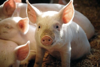
The Porcine Model: Gold Standard for Coronary Preclinical Studies
The pig’s cardiac anatomy and coronary physiology closely resemble those of humans, enabling clinical-like procedures with the same catheters, guidewires, and stents used in patients. This anatomical fidelity yields highly relevant data on vascular response and device performance. Pigs support high-quality angiography, intravascular ultrasound (IVUS), optical coherence tomography (OCT), and tissue harvesting for histology.
For these reasons, the porcine model is widely accepted — and often preferred — by regulators for coronary safety assessments. Published consensus recommendations on preclinical DES evaluation reinforce its central role in the development pipeline.
- Coronary artery diameter comparable to adult humans
- Compatible with full clinical catheterization equipment
- Supports multimodal imaging: angiography, IVUS, OCT
- Consistent neointimal response enabling reproducible studies
- Regulatory acceptance across FDA, EMA, and ISO frameworks
Inside a Stent Implantation Pig Model: Steps, Monitoring, and Deliverables

A typical stent implantation pig model protocol begins with veterinary screening and animal preparation, followed by femoral or carotid access, selective catheterization of the coronary arteries, baseline angiography, and stent deployment under fluoroscopic guidance. Antiplatelet and anticoagulation regimens mirror clinical practice. Follow-up imaging is scheduled at predefined timepoints, culminating in necropsy, perfusion fixation, and meticulous tissue collection for histopathology.
Deliverables generally include detailed procedural reports, baseline and follow-up angiograms with quantitative lumen measurements, intravascular imaging datasets, histomorphometry, cross-sectional images, and structured scoring for inflammation, fibrin deposition, and endothelial coverage. This integrated package is what transforms a study from an experiment into regulatory-grade evidence.
Common Pitfalls and Mitigation Strategies
- Incorrect stent-to-artery sizing — inflate injury scores and confound healing assessment
- Overexpansion-induced injury — non-uniform vessel trauma producing non-interpretable data
- Stent malapposition — mitigated via predefined acceptance criteria and consistent imaging
- Inconsistent procedural execution — addressed by standardized operating procedures and well-trained teams
The Rabbit Iliac Artery Model: When Smaller Is Smarter
The rabbit iliac artery model offers a cost-effective, accessible platform for comparative studies. Its paired design — where each animal serves as its own control — reduces variability and animal numbers while delivering high-quality histological outcomes. It is particularly useful for comparing coatings, drug formulations, strut thicknesses, and their effect on neointimal formation.
Limitations include vessel characteristics that differ from human coronaries and reduced compatibility with full-scale clinical equipment. For early screening and mechanistic comparisons, however, the rabbit iliac model remains a workhorse in any serious vascular stent animal study portfolio.
- Cost-effective paired design
- Each animal is its own control
- High-quality histological outcomes
- Ideal for early-stage screening
- Vessel characteristics differ from human coronaries
- Reduced compatibility with clinical equipment
- Does not replace porcine for pivotal coronary safety data
Healthy vs. Atherosclerotic Models: A Practical Comparison
Healthy models predominantly evaluate basic injury response, healing kinetics, and device-vessel interaction. Atherosclerotic models attempt to mimic human pathology, providing additional biological relevance for late-stage validation. The trade-off is real: disease models are more complex, more expensive, and sometimes less reproducible. Regulators typically accept standard healthy animal models for pivotal safety assessment, while disease models add value when specific pathophysiological questions must be answered.
| Aspect | Healthy Model | Atherosclerotic Model |
|---|---|---|
| Primary use | Injury response, basic healing | Disease-mimicking biology |
| Reproducibility | High | Moderate, variable |
| Cost & duration | Lower | Higher, longer induction |
| Regulatory acceptance | Standard pivotal use | Supportive / mechanistic |
| Endpoint clarity | Strong | Context-dependent |
Essential Endpoints in a Vascular Stent Animal Study
Endpoints fall into two categories: safety (thrombosis, inflammation, vessel injury) and performance (patency, neointimal area, late lumen loss). These are corroborated by histology and imaging. Histomorphometry quantifies neointimal area, percent stenosis, and injury scores. Healing assessment evaluates endothelial coverage, fibrin deposition, and macrophage presence. Device assessment confirms apposition, fracture, and — for DES — coating integrity. Insights from comparative design and manufacturing reviews reinforce why endpoint design must match stent technology.
Endpoint Specifics by Stent Type
For DES, special attention is paid to delayed healing, fibrin persistence, coating-related inflammation, and drug pharmacokinetics. For bioresorbable scaffolds, see recent advances in bioresorbable stent evaluation, which highlight degradation timelines and chronic vessel remodeling as primary endpoints.
- Thrombosis signals and patency
- Inflammation scoring
- Vessel injury and fibrin deposition
- Neointimal area and percent stenosis
- Late lumen loss and stent apposition
- Endothelial coverage and healing score
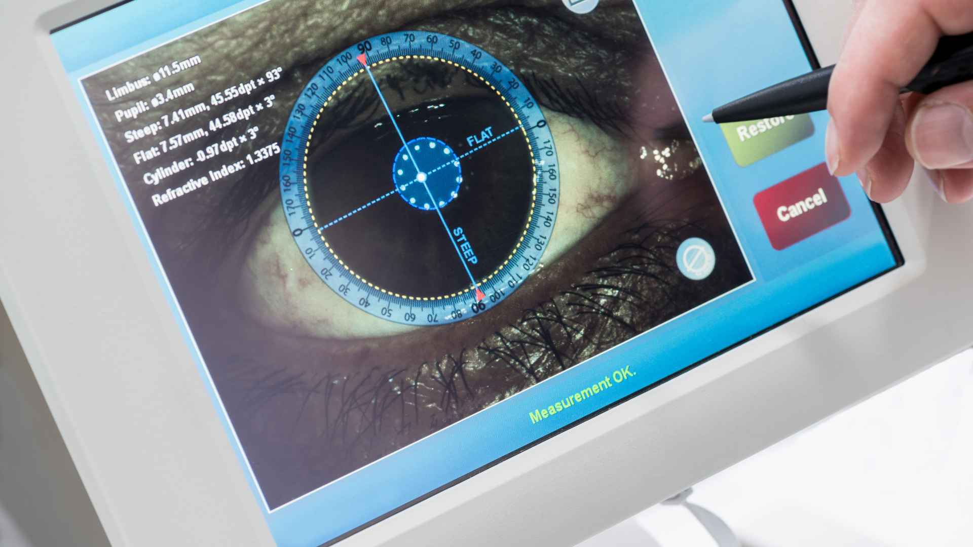
Imaging Modalities: Angiography, IVUS, and OCT
Angiography provides flow context and quantitative stenosis measurements. IVUS excels at volumetric assessment, vessel wall composition, and stent apposition over longer segments. OCT, with its micrometer-scale resolution, is the modality of choice for visualizing strut coverage, neointimal thickness, and early healing patterns.
Pivotal trial designs such as the SPIRIT FIRST trial of the XIENCE V stent reflect how preclinical imaging endpoints translate into clinical evidence. A robust coronary stent preclinical program rarely relies on a single modality; multimodal imaging plus histology delivers the most defensible dataset.
Our facility is equipped with C-Arm fluoroscopy, high-definition ultrasound, cardiac echocardiography, OCT, and surgical microscopy — the same imaging environment used in clinical practice — ensuring preclinical data directly mirrors clinical imaging standards.
Follow-up Durations: 28, 90, 180 Days and Beyond
Follow-up timing is dictated by the stent’s risk profile and mechanism of action. A 28-day window captures early injury response and acute healing. Ninety to 180 days assesses late healing, sustained drug effects, and neointimal maturation. Bioresorbable scaffolds may require 12 months or more to fully characterize degradation and vessel restoration. Choosing the right timepoints up front avoids costly study extensions later.
| Timepoint | Primary Insight | Typical Use |
|---|---|---|
| 7–14 days | Acute injury, thrombus | Safety screening |
| 28 days | Early healing, neointima onset | BMS baseline |
| 90 days | Mature neointima | DES safety |
| 180 days | Late healing, late catch-up | DES pivotal |
| 365+ days | Degradation, remodeling | Bioresorbable scaffolds |
Sample Size Planning for Preclinical Stent Studies
Sample size is driven by statistical power, model variability, and regulatory expectations. For large animal studies, group sizes of 8–12 animals per arm per timepoint are common, though specific targets depend on the primary endpoint and expected effect size. Careful planning aligned with ARRIVE guidelines ensures sufficient power to detect meaningful differences without unnecessary animal use — a balance that protects both scientific integrity and ethical standards.
At Biotech Farm, every study begins with a power calculation review. Our biostatisticians work alongside the veterinary team to define group sizes that satisfy both regulatory requirements (ISO 25539-2, FDA guidance) and ARRIVE 2.0 reporting standards, minimizing animal use while maximizing data quality.
Ethical Considerations and the 3Rs Framework
Every animal study must adhere to strict ethical guidelines. The 3Rs — Replacement, Reduction, Refinement — guide protocol design from the earliest planning stage. Institutional animal care and use committees (IACUCs) or equivalent ethics bodies must approve protocols before any procedure begins. Transparent reporting, comprehensive animal welfare monitoring, and skilled veterinary oversight are non-negotiable.
Facilities that combine professional crews with high tender care for the animals deliver not only better ethics but, in our experience, more consistent biological data. Animal welfare is not a constraint on good science — it is a prerequisite for it.
Use alternative methods wherever scientifically valid to reduce reliance on in vivo studies.
Minimize animal numbers through rigorous statistical planning and paired or crossover designs.
Continuously improve procedures to minimize suffering and maximize welfare through attentive veterinary care.
Regulatory Standards: ISO, FDA, and EMA
Stent testing is shaped by international standards and regional regulatory guidance. ISO 25539-2 covers endovascular devices including preclinical in vivo evaluation expectations. FDA guidance details non-clinical engineering tests and recommended labeling for intravascular stents. EMA guidelines address clinical and non-clinical evaluation, particularly for drug-eluting stents incorporating an ancillary medicinal substance.
A current view of regenerative principles applied to vascular stent design illustrates how technology evolution continuously reshapes regulatory expectations. Understanding these frameworks early in development prevents costly redesigns during submission.
| Standard / Body | Scope | Key Focus Areas |
|---|---|---|
| ISO 25539-2 | Endovascular devices | Preclinical in vivo evaluation, biocompatibility |
| FDA Guidance | Intravascular stents | Non-clinical engineering tests, labeling requirements |
| EMA Guidelines | DES with medicinal substance | Clinical and non-clinical evaluation for combination devices |
Good Laboratory Practice (GLP): The Quality Backbone

GLP is a quality system ensuring consistency, reliability, integrity, and reproducibility of non-clinical safety studies. It governs organization, personnel qualifications, facilities, equipment, test systems, study conduct, quality assurance, and reporting. A GLP stent animal study is not merely a study performed in a compliant facility — it is a study designed, executed, monitored, and documented to a standard that regulators can independently audit.
For pivotal submissions, GLP is essentially the price of entry. Every deviation, every observation, and every raw data point must be traceable, auditable, and defensible. This is why experienced facility management and transparent documentation culture matter as much as surgical skill.
- Qualified Study Director with documented responsibility
- Validated equipment with current calibration records
- Independent Quality Assurance inspection of critical phases
- Complete raw data retention with audit trail
- Final report signed by Study Director with QA statement
Histomorphometry: The Definitive Biological Readout
Histomorphometry provides quantitative analysis of tissue sections from stented vessels. Key measurements include neointimal area, percent area stenosis, injury score, inflammation score, fibrin score, and endothelial coverage. Performed by experienced pathologists using validated scoring systems, histomorphometry remains the ultimate method for evaluating long-term biological interaction between the stent and the vessel wall.
It is the endpoint that ties everything — imaging, pharmacology, mechanics — back to biology. No imaging modality alone can replace the cellular-level insight delivered by high-quality histopathology performed by specialists with deep anatomical understanding.
“Deep anatomical understanding is not a background qualification — it is the active lens through which every tissue section is interpreted and every regulatory question is answered.”
— Adir Koreh, Biotech Farm Ltd. & Biotech Anatomy Ltd.
Assessing Thrombosis Risk Preclinically
Thrombosis risk assessment combines acute and sub-acute observation, gross pathology at necropsy, and detailed histological examination for fibrin deposition and thrombus formation. Specific scoring systems quantify thrombus burden and vessel patency. Pharmacological protocols — particularly dual antiplatelet therapy — mirror clinical scenarios, allowing meaningful translation to expected patient management.
Variability in healing response across animals also informs labeling around minimum antiplatelet duration. This direct link between preclinical data and clinical prescribing guidance is one of the most impactful contributions of a well-designed thrombosis assessment protocol.
Common Mistakes That Undermine Preclinical Stent Programs
Several recurring missteps compromise otherwise promising programs. Each is avoidable with disciplined planning, predefined acceptance criteria, and an experienced preclinical team.
- Oversizing the stent to the artery — inflates injury scores and confounds healing assessment
- Mixing too many variables in a single study reduces statistical power
- Inadequate imaging schedules — miss critical healing windows
- Insufficient histology sampling — yields fragmentary biological insight
- Weak chain of custody for tissue specimens — jeopardizes data integrity
- No predefined acceptance criteria — decisions made retrospectively undermine regulatory credibility
The Role of a Specialized Stent Testing CRO
Contract research organizations dedicated to stent testing animal models provide the combination of expertise, facilities, and regulatory fluency that internal teams rarely maintain in-house. Services span study design, protocol development, GLP-compliant execution, advanced imaging, histopathology, statistical analysis, and regulatory reporting.
A capable stent testing animal models CRO helps device developers navigate FDA, EMA, and ISO expectations while compressing timelines — turning regulatory complexity into a manageable workflow rather than a bottleneck. The key differentiator is not simply access to animals, but the depth of scientific and procedural expertise brought to each program.
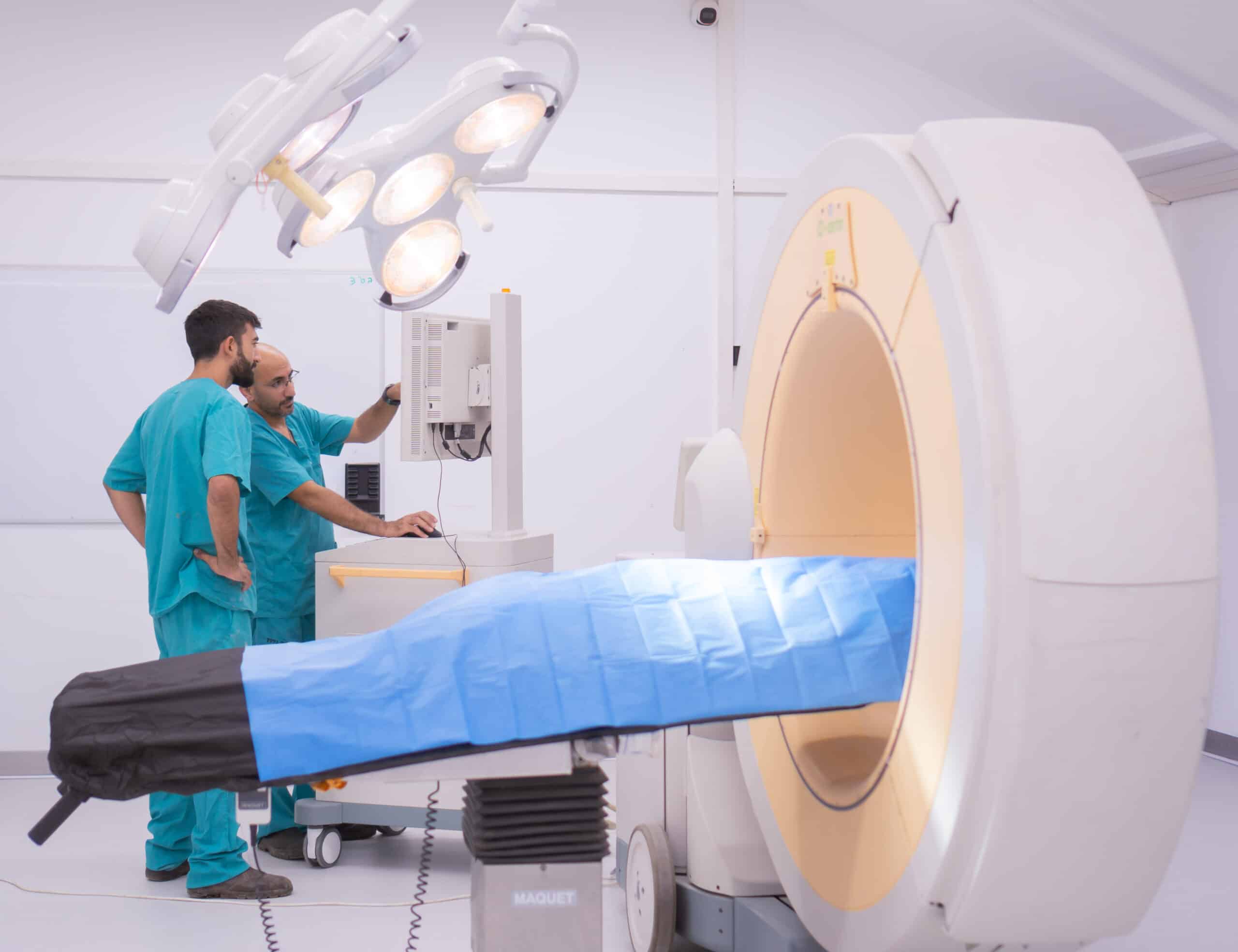
How Biotech Farm Supports Preclinical Stent Programs
Biotech Farm operates as a state-of-the-art preclinical facility built around three decades of leadership in large animal research. Our surgical suites are equipped with C-Arm fluoroscopy, high-definition ultrasound, cardiac echocardiography, OCT, and surgical microscopy — the same imaging environment used in clinical practice. Senior surgeons, experienced veterinary staff, and dedicated study managers collaborate on each program, with transparency in documentation as a core value rather than an afterthought.
Our porcine, ovine, and rabbit platforms cover the full spectrum of coronary stent preclinical, peripheral, and drug-eluting stent testing needs. Studies are conducted with attention to animal welfare, regulatory expectations, and scientific rigor. From a tailored study design to GLP-quality reporting, the goal is to deliver data that holds up to both scientific review and regulatory scrutiny.
| Business Need | How Biotech Farm Helps in Practice |
|---|---|
| Clinical-like procedures | Human-sized catheters and full interventional imaging suite |
| Multi-endpoint data | Integrated angiography, OCT, IVUS, and histomorphometry |
| Regulatory submission readiness | GLP-aligned workflows and structured reporting |
| Flexible study design | Tailored approach matching scope, timeline, and budget |
| Ethical execution | 3Rs framework, IACUC oversight, attentive animal care |
| Scientific partnership | Senior team available for protocol design and interpretation |
Frequently Asked Questions
Ready to Plan Your Next Preclinical Stent Study?
Which stent technology, indication, and timeline are you working toward — and how can your preclinical strategy be aligned to deliver the strongest possible evidence package? Our team is available to discuss study design, model selection, imaging endpoints, and GLP requirements tailored to your program.