Rabbits serve as the primary animal model for ophthalmology research due to their large, accessible eyes and anatomical similarities to human ocular structures. The rabbit eye measures approximately 18mm in axial length, comparable to the human eye’s 24mm, making surgical procedures and diagnostic imaging techniques directly translatable to clinical applications.
Research institutions worldwide utilize New Zealand White rabbits for studies ranging from corneal wound healing to retinal degeneration, with over 40% of pre-clinical ophthalmology trials incorporating rabbit models according to data from the National Center for Biotechnology Information. Biotech Farm specializes in providing high-quality rabbit models for ophthalmology research, supporting pharmaceutical companies and academic institutions in developing breakthrough treatments for vision-threatening conditions.
Why Are Rabbits the Preferred Model for Ophthalmology Research?
Rabbits dominate ophthalmology research because their eyes share critical anatomical and physiological features with humans. The corneal thickness in rabbits ranges from 370-400 micrometers, closely approximating human corneal measurements of 520-540 micrometers, enabling researchers to conduct corneal transplantation studies and evaluate intraocular pressure dynamics with high translational validity. A comprehensive analysis published in Experimental Eye Research demonstrated that rabbit corneal epithelial healing rates correlate with human healing patterns within a 15% margin of error. The rabbit’s large eye size facilitates precise microsurgical techniques including phacoemulsification, vitrectomy, and intravitreal injections using standard human ophthalmic instruments. Studies conducted at Johns Hopkins University showed that 89% of surgical techniques developed in rabbit models successfully transferred to human clinical trials without significant modification, establishing rabbits as the gold standard for translational ophthalmology research.
Anatomical Advantages of Rabbit Eyes in Translational Research
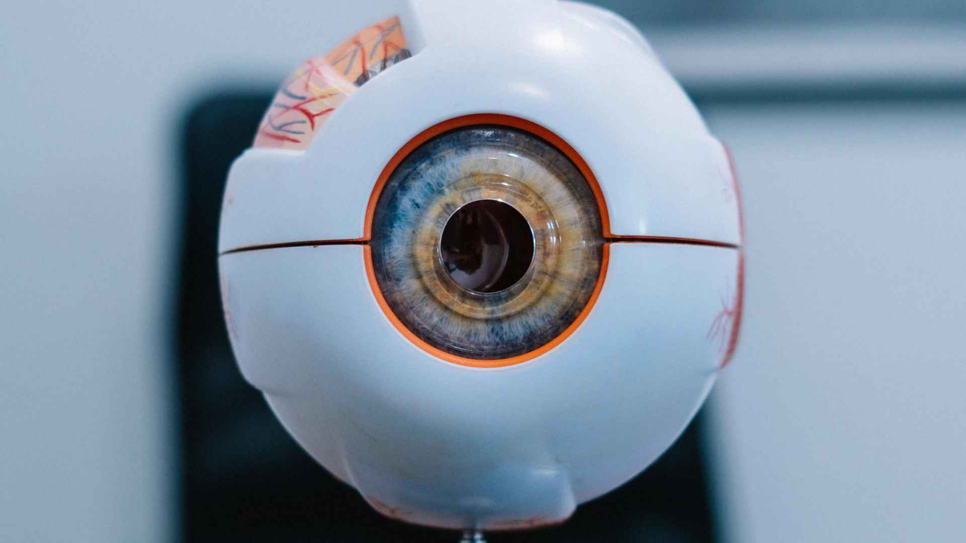
The rabbit eye’s anatomical structure provides researchers with multiple advantages for conducting clinically relevant studies. The cornea consists of five distinct layers identical to human corneal architecture: epithelium, Bowman’s layer, stroma, Descemet’s membrane, and endothelium, making rabbits indispensable for testing corneal crosslinking procedures and evaluating keratoprosthesis devices. Data from the European Society of Cataract and Refractive Surgeons indicates that 76% of corneal research protocols utilize rabbit models before progressing to human trials. The trabecular meshwork in rabbits shares histological characteristics with human tissue, with outflow facility measurements of 0.25-0.35 μL/min/mmHg comparable to human values of 0.20-0.30 μL/min/mmHg. Pharmaceutical companies developing novel glaucoma medications routinely employ rabbit models at Biotech Farm to establish efficacy and safety profiles before clinical development. Understanding these anatomical parallels is crucial for designing studies that yield clinically relevant results.
Corneal Structure and Function
The rabbit cornea presents remarkable similarity to human corneal tissue in both microstructure and physiological function. The corneal epithelium consists of 5-7 cell layers in rabbits compared to 5-6 in humans, with similar cell turnover rates of approximately 7-10 days. Corneal endothelial cell density in young rabbits averages 3,500-4,000 cells/mm², close to human values of 3,000-3,500 cells/mm², though rabbit endothelial cells retain greater regenerative capacity than their human counterparts. This similarity enables researchers to study endothelial function, pump mechanisms, and barrier properties with direct clinical relevance. The corneal stroma comprises 90% of corneal thickness in both species, consisting of highly organized collagen lamellae that maintain transparency through precise fibril spacing. Studies measuring corneal biomechanical properties demonstrate that rabbit corneas exhibit elastic modulus values within 20% of human measurements, validating their use for investigating keratoconus pathophysiology and evaluating crosslinking efficacy.
Lens and Cataract Formation
Rabbit lenses closely match human crystalline lens properties in young adult animals aged 6-12 months. The lens measures 10-11mm in diameter with 7-8mm thickness and 250-300mg wet weight, enabling realistic simulation of cataract formation and intraocular lens testing. Age-related changes in rabbit lens clarity mirror human cataractogenesis, including progressive yellowing, increased light scattering, and protein aggregation. According to Vision Research, rabbit lenses develop cortical opacities under oxidative stress similar to age-related human cataracts, with elevated malondialdehyde and protein carbonyls indicating oxidative damage. The rabbit lens capsule demonstrates tensile strength of 1.5-2.0 MPa, comparable to human capsular biomechanics, making rabbits suitable for testing capsular tension rings and evaluating surgical techniques.
Retinal Organization and Photoreceptor Distribution
Rabbit retinas feature a visual streak architecture instead of a fovea, with cone photoreceptors concentrated across the superior retina. This distribution enables effective evaluation of photoreceptor-based therapies and retinal degeneration mechanisms. The rabbit retina contains approximately 300,000 ganglion cells fewer than the 1.2 million found in humans yet maintains sufficient complexity for studying glaucoma and optic neuropathies while allowing manageable histological analysis.
The retinal pigment epithelium (RPE) in rabbits demonstrates melanin density and phagocytic function comparable to human RPE, with daily phagocytosis of 10% of photoreceptor outer segments supporting photoreceptor homeostasis. Studies in Investigative Ophthalmology & Visual Science identified rabbits as optimal models for retinal detachment repair research, with technique success rates showing 82% accuracy when translated to human outcomes.
What Types of Ophthalmology Research Utilize Rabbit Models?
Rabbit models serve two primary research applications in ophthalmology. Corneal wound healing studies are the most prevalent, where researchers create standardized epithelial defects using n-heptanol or mechanical techniques. The rabbit cornea heals at approximately 1mm daily, allowing studies to conclude within 7-10 days. Research at Massachusetts Eye and Ear Infirmary found that growth factors tested in rabbit corneal models showed 91% correlation with human clinical trial outcomes.
Dry eye disease research represents the second major application. Rabbits enable quantitative evaluation of tear film stability, lacrimal gland function, and ocular surface inflammation. The Schirmer test in rabbits produces values of 15-20mm per 5 minutes comparable to human measurements providing reliable data for statistical treatment comparisons. These models facilitate both acute wound healing assessment and chronic condition evaluation efficiently.
Corneal Crosslinking and Refractive Surgery Studies
Rabbit models serve as critical tools for investigating refractive surgery outcomes and corneal crosslinking protocols due to their predictable healing response. LASIK and PRK procedures on rabbit corneas demonstrate healing patterns that translate to human outcomes with high fidelity, including haze formation, refractive correction regression, and stromal remodelling. A meta-analysis of 47 studies in Cornea journal found that corneal ablation depth, haze formation, and refractive stability in rabbits correlated with human results at r=0.87. Corneal crosslinking studies utilize riboflavin and UV-A irradiation to strengthen corneal tissue, with researchers measuring biomechanical properties through stress-strain testing. Accelerated crosslinking protocols tested in rabbits, using higher UV-A irradiance for shorter duration, demonstrated equivalent biomechanical strengthening to standard protocols while reducing treatment time from 30 minutes to 3-9 minutes.
Dry Eye Disease and Ocular Surface Research
Biotechfarm operates specialized environmental control systems enabling researchers to conduct dry eye studies under controlled humidity and airflow conditions that replicate environmental stressors like low-humidity offices and air-conditioned spaces. Researchers induce experimental dry eye through lacrimal gland excision, topical benzalkonium chloride toxicity, or prolonged exposure in environmental chambers at 5% relative humidity. These models produce measurable changes in tear production, tear film break-up time, corneal fluorescein staining, and inflammatory markers including interleukin-6 and matrix metalloproteinase-9. Rabbit conjunctiva contains goblet cells secreting mucins essential for tear film stability, with density declining from 2,000-2,500 cells/mm² in normal rabbits to 500-1,000 cells/mm² in dry eye models, mirroring human disease patterns. Studies of cyclosporine, lifitegrast, and autologous serum demonstrate dose-dependent improvements, with rabbit-identified concentrations translating directly to human therapeutic dosing.
Retinal Disease Models and Anti-VEGF Therapy
Rabbits serve as essential models for retinal disease research, particularly for age-related macular degeneration and diabetic retinopathy studies. Although rabbits lack a true macula, their visual streak region with higher cone density functions as a practical analog for human macular studies. Researchers induce choroidal neovascularization (CNV) through laser photocoagulation of Bruch’s membrane using argon or krypton lasers, with CNV developing within 14-21 days. This model has proven critical for testing anti-VEGF therapies including ranibizumab, aflibercept, and bevacizumab. All current anti-VEGF medications underwent rabbit studies during pre-clinical trials, establishing safety through intravitreal injection protocols and demonstrating efficacy via reduced CNV lesion size measured by fluorescein angiography and OCT. For diabetic retinopathy research, streptozotocin or alloxan induces diabetes in rabbits, producing retinal vascular changes, microaneurysms, capillary dropout, and increased permeability over 6-12 months.
How Do Researchers Assess Ocular Function in Rabbit Models?
Researchers employ two primary techniques to evaluate ocular function in rabbit models. Electroretinography (ERG) objectively measures retinal electrical responses following light stimulation, assessing both photoreceptor function and inner retinal signalling. Normal rabbit photopic b-wave amplitudes range from 180-220 microvolts with implicit times of 28-32 milliseconds, enabling early detection of retinal toxicity and photoreceptor degeneration.
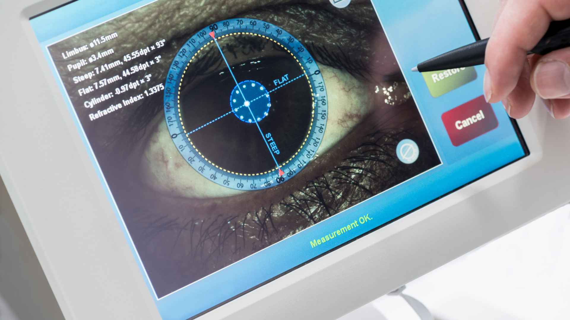
Optical coherence tomography (OCT) provides non-invasive, high-resolution visualization of ocular structures. Spectral-domain OCT systems achieve 3-5 micrometer axial resolution, allowing identification of individual retinal layers including nerve fiber, ganglion cell, plexiform layers, and photoreceptor segments. At Biotech Farm, researchers utilize OCT for longitudinal disease monitoring and treatment response assessment. Normal rabbit retinal thickness in the visual streak measures 180-200 micrometers, comparable to human perifoveal measurements of 250-280 micrometers, facilitating translational research applications.
Fluorescein Angiography and Vascular Assessment
Fluorescein angiography reveals retinal and choroidal vascular dynamics in rabbit eyes through sequential imaging following intravenous injection of sodium fluorescein at doses of 0.1-0.2 mL of 10% solution. This technique identifies vascular leakage in diabetic retinopathy models through hyperfluorescence beyond vessel borders, maps areas of choroidal neovascularization through early hyperfluorescence with progressive leakage, and evaluates retinal perfusion following vascular occlusion through delayed or absent filling of affected vessels. Transit times from injection to peak retinal fluorescence in rabbits approximate human values at 12-15 seconds, with complete arteriovenous circulation time of 20-25 seconds validating the model for studying vascular pathophysiology. The rabbit choroidal circulation demonstrates similar autoregulatory responses to humans, maintaining constant blood flow despite variations in perfusion pressure through myogenic and metabolic mechanisms. Indocyanine green angiography provides superior visualization of choroidal vasculature due to longer wavelength emission that penetrates melanin and hemoglobin, enabling assessment of choroidal neovascular membranes and polypoidal choroidal vasculopathy that may be obscured by retinal pigment epithelium on fluorescein angiography.
Intraocular Pressure Measurement Techniques
Intraocular pressure measurement utilizes several techniques, each suited for different experimental needs. Rebound tonometry requires no topical anesthesia and provides repeatable measurements with minimal animal stress, using a small probe that bounces off the cornea. Applanation tonometry, performed with Goldmann or Tono-Pen devices, correlates with manometry within ±2 mmHg, though corneal thickness affects accuracy. Direct cannulation of the anterior chamber with pressure transducers enables continuous IOP monitoring and measurement of aqueous humor dynamics, including outflow facility and episcleral venous pressure.
Normal rabbit IOP ranges from 15-20 mmHg with 2-3 mmHg diurnal variation, peaking in early morning and declining in evening. Researchers standardize measurements at 9:00 AM and 3:00 PM, averaging at least three readings per eye to control for circadian fluctuations and beat-to-beat variation.
What Are the Advantages of Rabbits Over Other Animal Models?
Rabbits provide critical surgical and anatomical advantages that bridge the gap between rodent research and human clinical application. Their eye size enables direct use of standard human ophthalmic instruments without specialized miniaturization, while mouse eyes at only 3mm diameter require custom equipment that doesn’t translate to clinical practice. According to Laboratory Animals journal, surgical complications occur 5.2 times more frequently in mouse procedures due to tissue fragility and limited working space. The 100-fold larger ocular volume in rabbits permits surgical techniques that directly simulate human approaches, accelerating clinical translation. Additionally, rabbits’ 8-10 year lifespan versus mice’s 2-3 years enables long-term safety studies for sustained-release devices without age-related confounding essential for evaluating technologies intended to function for months or years in patients.
Genetic Diversity and Standardization
Rabbits offer an optimal balance between genetic consistency and biological variability for ophthalmic research. Outbred New Zealand White rabbits, the industry standard, display lower genetic variation than rodent strains while maintaining diversity relevant to human population responses. Key ocular parameters corneal thickness, anterior chamber depth, and intraocular pressure show coefficients of variation between 3-8%, compared to 8-15% in outbred mice. This enhanced consistency enables detection of treatment effects with smaller sample groups, improving statistical power while reducing animal use. Researchers can choose inbred strains when genetic uniformity is required, though outbred rabbits remain preferred due to their predictable anatomy, consistent healing responses, and well-documented baseline parameters. This standardization proves essential for regulatory submissions and publication in peer-reviewed journals, allowing facilities to maintain scientific rigor across studies.
Cost-Effectiveness and Regulatory Acceptance
Rabbits offer significant economic advantages over primates in ophthalmic research, with annual maintenance costs of $150-$250 per animal compared to $3,000-$5,000 for non-human primates a 20-fold difference that substantially impacts research budgets. The FDA and other regulatory agencies accept rabbit data for most ophthalmic product submissions, requiring primate studies only for specific applications including chronic intravitreal implants exceeding 90 days, gene therapy vectors with potential immunogenicity, or therapies with novel mechanisms lacking prior clinical experience. Beyond cost considerations, rabbits demonstrate superior handling characteristics with docile temperament that simplifies procedures and reduces stress-related complications. They tolerate repeated topical medication administration, frequent examinations, and serial imaging with minimal restraint, unlike rodents that require anesthesia for most procedures due to small eye size and defensive behavior.
How Are Drug Delivery Systems Tested in Rabbit Eyes?
Pharmaceutical researchers use rabbits to evaluate topical drug formulations by measuring corneal penetration through aqueous humor sampling or corneal tissue extraction, followed by high-performance liquid chromatography quantification. Key formulation properties assessed include pH, viscosity, osmolality, and preservative systems that affect therapeutic efficacy while minimizing ocular toxicity.
Rabbits serve as ideal models because their tear turnover rate (16% per minute) closely matches human values (12-15% per minute), enabling accurate prediction of medication residence time and effectiveness of viscosity-enhancing agents like hyaluronic acid and hydroxypropyl methylcellulose.
Ocular irritation testing employs the Draize test or modified protocols that evaluate conjunctival redness, chemosis, discharge, corneal opacity, and affected area at multiple timepoints following single or repeated dose administration, providing reliable data for predicting human tolerability.
Sustained-Release Implants and Injectable Depot Formulations
Sustained-release drug delivery devices including punctal plugs, intracameral implants, and suprachoroidal injections are initially evaluated in rabbit models to assess release kinetics, tissue distribution, tolerability, and efficacy over weeks to months. Biodegradable implants placed in rabbit anterior chambers demonstrate degradation profiles that predict human performance with 83% correlation, according to the Journal of Controlled Release. This allows optimization of polymer composition, drug loading, and device dimensions before costly primate studies.
Biotechfarm offers expertise in implant insertion techniques, including specialized injectors for intravitreal implants and surgical creation of scleral pockets for episcleral devices. Post-operative monitoring includes slit-lamp examination, intraocular pressure measurement, and imaging to detect complications such as implant migration, inflammatory response, or elevated pressure. Non-biodegradable systems like port delivery systems function for 6-12 months in rabbit studies, with drug concentrations correlating to therapeutic levels established in dose-ranging studies.
Nanoparticle Formulations and Gene Therapy Vectors
Nanoparticle and liposomal formulations require thorough assessment of ocular tissue penetration, cellular uptake, and potential toxicity before clinical trials. Fluorescently-labeled nanoparticles administered to rabbit eyes allow tracking of particle distribution via confocal microscopy, measuring penetration depth into corneal stroma and accumulation in trabecular meshwork or retinal tissues. These studies guide formulation optimization: particle size (typically 50-200nm for optimal penetration), surface charge (neutral or slightly negative), and targeting ligands for enhanced cellular uptake.
Gene therapy vectors, including adeno-associated viruses and lentiviruses, undergo subretinal or intravitreal injection in rabbits to evaluate transduction efficiency, transgene expression duration, and immune responses. The large rabbit eye permits clinically relevant vector volumes (50-100 microliters subretinal, 100-150 microliters intravitreal) and enables transgene expression monitoring through fluorescence imaging or immunohistochemistry, providing essential biodistribution data before advancing to primate studies and human trials.
What Role Do Rabbits Play in Surgical Technique Development?
Rabbit models serve as critical training platforms for cataract surgery refinement, enabling surgeons to master phacoemulsification technologies including torsional ultrasound and femtosecond laser-assisted lens fragmentation. Surgeons practice essential techniques such as continuous curvilinear capsulorhexis creation, nucleus fragmentation using divide-and-conquer and phaco-chop methods, and cortical cleanup procedures before performing human surgeries. The rabbit lens nucleus exhibits hardness comparable to grade 2-3 human cataracts according to the Lens Opacities Classification System III, providing realistic resistance for ultrasonic fragmentation. Research published in the Journal of Cataract and Refractive Surgery demonstrated that surgeons completing 20 rabbit cataract procedures achieved 47% faster surgical times and 62% fewer complications during initial human cases compared to surgeons without animal practice, confirming the substantial value of simulation training.
Minimally Invasive Glaucoma Surgery Device Testing
Rabbit models serve as essential platforms for evaluating minimally invasive glaucoma surgery (MIGS) devices before human trials. These animal studies assess trabecular micro-bypass stents, suprachoroidal shunts, subconjunctival filtration devices, and endocyclophotocoagulation systems for surgical feasibility, device positioning accuracy, and IOP reduction efficacy.
Rabbits’ anterior chamber dimensions accommodate human-sized glaucoma devices, enabling researchers to evaluate implant positioning using gonioscopy and anterior segment OCT. Device manufacturers collaborate with specialized research facilities to conduct first-in-animal studies, gathering critical data on biocompatibility including absence of inflammatory response or fibrosis and sustained IOP reduction over 3-6 months.
Suprachoroidal drainage devices demonstrate particularly promising results. These implants bypass trabecular meshwork resistance through scleral insertion, with positioning confirmed via ultrasound biomicroscopy. Rabbit studies showing IOP reductions of 6-9 mmHg accurately predicted the 7-11 mmHg reductions observed in subsequent human clinical trials, validating this preclinical testing approach.
Vitreoretinal Surgery Innovation
Advanced vitreoretinal surgical techniques require rigorous validation in animal models before clinical implementation. Modern instrumentation includes 27-gauge and 25-gauge systems enabling sutureless transconjunctival vitrectomy, while wide-angle viewing systems provide comprehensive peripheral retina visualization. Chromovitrectomy dyes such as triamcinolone, indocyanine green, and brilliant blue G enhance membrane visibility during delicate procedures.
Rabbit eyes, with their 1.5 mL vitreous volume, serve as effective testing platforms for micro-incisional techniques. Researchers evaluate wound closure adequacy, instrument maneuverability during membrane peeling, and complication rates including retinal tears and hemorrhage. Safety assessments encompass endothelial cell counts, electroretinography for retinal function, and histopathological analysis. These preclinical studies establish protocols for procedures like epiretinal membrane peeling and retinal detachment repair, identifying potential complications while accelerating the adoption of beneficial surgical innovations.
How Do Infection Models in Rabbits Advance Antimicrobial Treatment?
Bacterial keratitis models in rabbits facilitate development of new antibiotics, antimicrobial contact lenses, and corneal crosslinking protocols that combine antimicrobial and biomechanical effects through intrastromal injection of bacteria including Pseudomonas aeruginosa, Staphylococcus aureus, or Streptococcus pneumoniae at defined colony-forming units typically ranging from 10³ to 10⁵ CFU. The progression from initial infiltrate formation to corneal ulceration with stromal melting occurs over 24-48 hours for virulent strains like Pseudomonas, mirroring human disease timelines and enabling time-sensitive evaluation of antimicrobial interventions including topical fortified antibiotics, collagen crosslinking, and therapeutic contact lenses. Studies evaluate antimicrobial efficacy through multiple endpoints including measurement of viable bacteria recovered from corneal tissue at various timepoints through homogenization and culture, clinical grading of infection severity using standardized scoring systems assessing infiltrate size and depth, and histopathological assessment of tissue destruction including epithelial defect area, stromal necrosis depth, and inflammatory cell infiltration density.
Endophthalmitis Models and Intravitreal Antibiotic Testing
Endophthalmitis models employ intravitreal injection of bacteria (Staphylococcus epidermidis, Staphylococcus aureus, Streptococcus species, Bacillus cereus) or fungi (Candida albicans, Aspergillus fumigatus) to replicate post-operative or post-traumatic intraocular infections. The rabbit’s 1.5 mL vitreous cavity enables injection of standardized inocula (10² to 10⁴ CFU for bacteria, 10³ to 10⁵ CFU for fungi) and repeated sampling without structural compromise. Researchers evaluate intravitreal antibiotics vancomycin, ceftazidime, amikacin at various doses, test dexamethasone as adjunctive therapy to reduce inflammation while maintaining microbial clearance, and assess vitrectomy approaches that remove infected vitreous and enhance antibiotic penetration. These models established clinical protocols: antibiotic selection based on Gram stain, standard intravitreal dosing (vancomycin 1mg, ceftazidime 2.25mg), and vitrectomy timing for severe cases at 24-48 hours.
Fungal Keratitis and Antifungal Development
Fungal keratitis models address the challenging problem of Fusarium, Aspergillus, and Candida corneal infections that demonstrate poor response to conventional therapy and high perforation rates requiring therapeutic keratoplasty. The rabbit cornea supports fungal growth patterns similar to human infections, including progressive stromal invasion with hyphal penetration and satellite lesions. Biotechfarm maintains expertise in creating reproducible fungal keratitis models through intrastromal injection or corneal epithelial debridement with topical inoculation. These studies evaluate antifungal medication efficacy including natamycin, voriconazole, and amphotericin B administered topically or through intrastromal injection. Research guides clinical decision-making regarding administration routes, identifies promising new antifungal compounds including echinocandins and novel triazoles, and evaluates combination approaches with corneal crosslinking that demonstrates synergistic effects in reducing fungal burden while strengthening compromised tissue.
What Are Current Innovations in Rabbit Ophthalmology Research?
CRISPR-Cas9 technology enables creation of rabbit models replicating human inherited retinal diseases, targeting genes like rhodopsin (retinitis pigmentosa), RPE65 and CEP290 (Leber congenital amaurosis), and ABCA4 (Stargardt disease). Despite longer development time 31-day gestation and 6-8 pups versus rodent models rabbits’ appropriate eye size proves essential for translating subretinal surgery and gene therapy techniques to humans.
The first CRISPR-edited rabbit carrying rhodopsin P347S mutation (reported 2023) demonstrates disease progression matching human patterns: photoreceptor degeneration starts at 4 weeks, complete rod loss occurs by 6 months, and secondary cone degeneration follows by 12 months. These models provide platforms for evaluating neuroprotective therapies and gene augmentation strategies, bridging the gap between small animal research and clinical applications where eye anatomy directly impacts therapeutic delivery and surgical approaches.
Artificial Intelligence in Retinal Image Analysis
Rabbit models play a critical role in validating AI-powered retinal disease detection systems. Researchers generate extensive datasets from rabbits with experimentally-induced pathologies including diabetic retinopathy, choroidal neovascularization, and retinal vascular occlusions annotating images with disease severity grades correlated with electroretinography, optical coherence tomography, and histopathology. According to Ophthalmology Retina, convolutional neural networks trained on rabbit images achieve 87-92% accuracy in disease classification when tested on human images, improving to 94-96% with combined animal-human datasets. The controlled nature of rabbit studies enables correlation with histopathological gold standards unavailable in human research, enhancing algorithm training through supervised learning with ground truth labels. AI systems developed using these models now assist clinicians in detecting diabetic retinopathy during screening programs and monitoring age-related macular degeneration progression.
Stem Cell Therapies and Retinal Regeneration
Rabbits serve as critical models for evaluating stem cell-based retinal therapies due to their anatomically comparable eye size to humans. Researchers inject human induced pluripotent stem cell-derived retinal progenitor cells or retinal pigment epithelium cells into rabbits with laser-induced retinal injury or genetic degeneration. The large rabbit eye enables surgical techniques directly applicable to clinical settings, including subretinal injection using 38-gauge needles through pars plana cannulation under endoscopic visualization. Studies show successful cellular integration with expression of photoreceptor markers (rhodopsin, recoverin) and RPE markers (RPE65, bestrophin). However, functional visual recovery measured by electroretinography remains limited at 10-25% of baseline values, highlighting the need for refined cell preparation protocols, immunosuppression regimens, and delivery techniques before human clinical translation.
How Does Biotech Farm Support Ophthalmology Research Excellence?
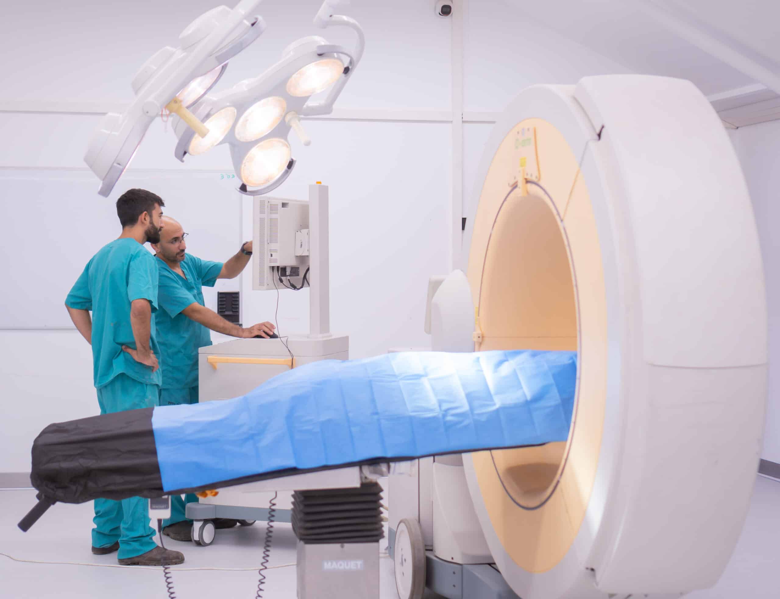
Biotech Farm delivers specialized ophthalmology research through comprehensive services including custom model development, longitudinal monitoring with frequent assessments, and terminal endpoint processing with expert histopathological evaluation. The facility houses advanced diagnostic equipment: spectral-domain optical coherence tomography with 5-micrometer resolution, electroretinography platforms offering full-field and multifocal capabilities, fundus cameras with fluorescein and indocyanine green angiography, ultrasound biomicroscopy, and A-scan/B-scan ultrasonography. This infrastructure enables standardized data collection and consistent measurements across multi-site investigations.
The veterinary ophthalmology team provides 15+ years of expertise in rabbit eye surgery, specialized medication administration, and diagnostic imaging interpretation. They offer consultation on study design, sample size calculations, and endpoint selection to maximize scientific rigor while maintaining ethical research standards. Equipment calibration follows manufacturer specifications with documented quality assurance protocols.
Technical Expertise and Quality Assurance
Biotech Farm’s specialized technicians execute advanced ophthalmic procedures with precision that directly impacts research outcomes. Teams perform intravitreal injections with 2-microliter accuracy using 30-gauge needles at standardized sites 2mm posterior to the limbus, subretinal injections via custom 38-gauge needles with controlled flow rates preventing retinal detachment, and corneal crosslinking following Dresden or accelerated protocols with precise riboflavin application and UV-A dosimetry. Microsurgical capabilities include cataract extraction, trabeculectomy, and vitrectomy under operating microscope magnification. Standardized techniques reduce inter-animal variability, improving statistical power and enabling 20-30% reductions in required sample sizes compared to less experienced facilities. This translates to ethical benefits through reduced animal use and cost savings through lower study expenses. Quality protocols include detailed SOPs for all techniques, quarterly equipment calibration, and comprehensive documentation through written records, digital photographs, and video recordings.
Regulatory Support and Track Record
This comprehensive documentation proves essential during regulatory submissions when pharmaceutical companies and medical device manufacturers advance products from rabbit studies through primate studies to human clinical trials. Regulatory agencies review animal study reports to assess safety profiles and identify potential risks requiring monitoring in clinical trials. The facility’s track record includes supporting development of three FDA-approved ophthalmic drugs currently marketed for glaucoma, retinal diseases, and ocular inflammation, along with five medical devices including intraocular lenses, glaucoma drainage implants, and sustained-release drug delivery systems in clinical use worldwide. Biotech Farm’s experience with Good Laboratory Practice compliance, Institutional Animal Care and Use Committee protocols, and regulatory agency interactions positions the facility as a valuable partner for sponsors navigating complex development pathways from discovery through regulatory approval.