Preclinical Imaging Services for Animal Studies: A Professional Guide to Planning, Execution, and Decision-Making
With over 30 years of combined expertise in preclinical research leadership and management, BIOTECH FARM Ltd. has guided hundreds of imaging studies from initial concept through regulatory-ready deliverables. Our team understands that preclinical imaging services represent far more than scanning animals — they form the quantitative backbone of critical go/no-go decisions that determine which therapeutic candidates advance to clinical trials.
At the heart of medical innovation, preclinical imaging services play a pivotal role in translating scientific discoveries into viable therapeutic candidates. These services provide comprehensive professional support for planning, executing, and analyzing in vivo and ex vivo imaging across animal models, enabling teams to assess efficacy, safety, and pharmacodynamics before clinical trials begin.
Expert Insight: Preclinical imaging enables non-invasive measurement of changes in tumor size, inflammation, vascular permeability, and pharmacodynamic markers over time. This makes it indispensable for biotech and pharmaceutical companies, academic researchers, and preclinical development teams who require standardized outputs and thorough documentation for regulatory submissions.
What Does In Vivo Imaging Actually Measure in Preclinical Research?
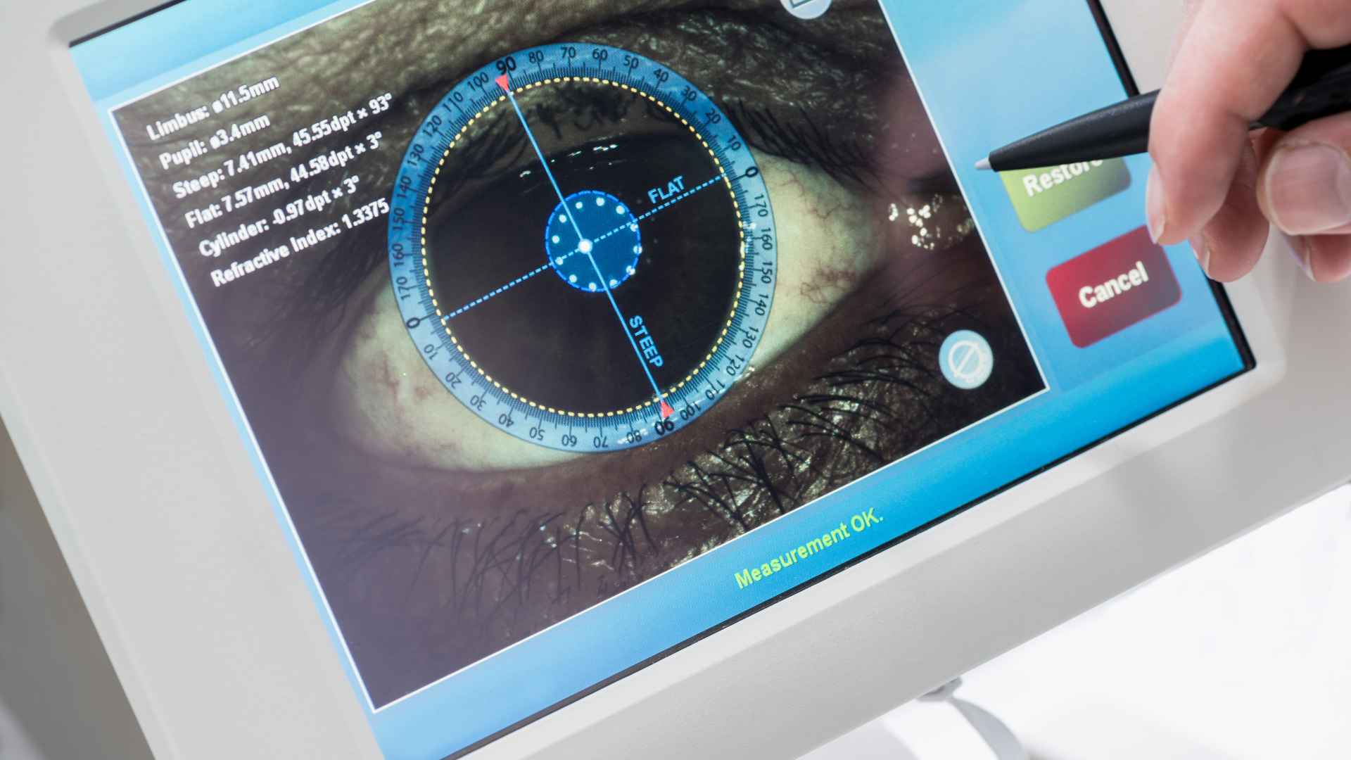
In vivo imaging involves scanning living animals to quantitatively assess anatomy, organ function, or molecular signals. Unlike terminal endpoint assays, it allows longitudinal monitoring of the same subject across multiple time points. Common measurements include:
- Tumor volume and growth kinetics
- Tissue perfusion and vascular permeability indices
- Tracer uptake in PET or SPECT studies
- Bioluminescence and fluorescence signal intensity
- Cardiac function and metabolic activity
The commercial advantage is substantial: fewer invasive endpoints, more data points per animal, and earlier go/no-go decision-making. When dynamic changes over time, treatment response assessment, or reduction of inter-animal variability are critical to the study objectives, in vivo imaging becomes preferable to relying on histology alone. Histology provides a single snapshot, while imaging captures the full trajectory of disease progression or therapeutic effect.
As described in detail at Preclinical Animal Studies: Key to Medical Breakthroughs, these studies represent a foundational step toward achieving regulatory approval and advancing patient care.
Choosing the Right Imaging Modality: PET, CT, MRI, Ultrasound, or Optical?
Modality selection depends on the biological question being asked, the tissue depth of interest, required spatial resolution, need for absolute quantification, availability of probes or tracers, and logistical factors such as scan duration and cost. A comprehensive review published in PubMed Central on multi-platform imaging in cancer models demonstrates how different modalities complement each other when addressing complex preclinical questions.

| Modality | Primary Strengths | Limitations | Typical Applications |
|---|---|---|---|
| Optical Imaging | High molecular sensitivity, real-time visualization | Limited tissue depth penetration | Bioluminescence, fluorescence reporter assays |
| MRI | Excellent soft tissue contrast, functional data | Longer scan times, higher cost | Neurology, oncology, cardiac function |
| PET/SPECT | High molecular sensitivity, quantitative tracer uptake | Requires radiochemistry infrastructure | Metabolic activity, receptor binding |
| CT | Fast acquisition, excellent bone/anatomy detail | Limited soft tissue contrast alone | Skeletal imaging, lung, combined with PET |
| Ultrasound | Fast, portable, real-time, cost-effective | Operator-dependent, limited field of view | Cardiac function, vascular flow, abdominal imaging |
Five Questions to Guide Your Modality Decision
Before selecting an imaging platform, answer these questions systematically to narrow the modality options and prevent costly protocol changes mid-study:
- What is the primary biological objective?
- What is the study timeframe and number of time points?
- What tissue depth must be reached?
- Is absolute quantification required for regulatory submissions?
- What are the budget constraints?
A Common Mistake: Confusing a Preclinical Imaging CRO with a Core Facility
A preclinical imaging CRO (Contract Research Organization) is a commercial service provider that manages imaging projects end-to-end. This includes project management, Service Level Agreements, quality assurance documentation, standardized acquisition protocols, and defined deliverables. A core facility, by contrast, typically operates on a shared-access or instrument-booking model where the researcher performs much of the work independently.
⚠️ Important Distinction: The CRO vs. core facility distinction matters most when facing tight deadlines, when inter-group comparisons require rigorous standardization, when quality documentation must satisfy regulatory reviewers, or when data needs to be transferred to multiple stakeholder teams. In these scenarios, a CRO provides the infrastructure and accountability that a core facility may not consistently deliver.
CRO Services Include:
- Dedicated project management
- Service Level Agreements
- Regulatory-ready documentation
- Standardized protocols
Core Facility Typically Offers:
- Instrument access booking
- Basic technical training
- Self-service model
- Variable documentation standards
What Actually Drives the Cost of Preclinical Imaging Services?
Costs are primarily determined by multiple interconnected factors that vary significantly between projects:
- Imaging modality selected — PET/SPECT requires radiochemistry infrastructure; MRI involves longer scan times
- Number of animals and time points — longitudinal studies cost more but reduce total animal numbers
- Complexity of image analysis — 3D segmentation and radiomics increase costs but improve decision quality
- Specialized probes, contrast agents, or radiotracers — custom synthesis adds significant expense
- Study design consultation and project management — comprehensive support vs. basic scanning services
???? Avoiding Surprises in Your Quote
Request an itemized breakdown that specifies:
- Cost per animal, per scan, and per analysis step
- Whether instrument time, animal preparation, anesthesia, and contrast agents are included
- Cost of protocol optimization runs versus production scans
- Final deliverables and report format specifications
Facilities like Biotech Farm, which integrate project management and scientific escort into their service model, help clients anticipate costs early by providing transparent, itemized quotes that break down each component clearly.
Realistic Timelines: From Scheduling Scans to Receiving Results

Lead time for most preclinical imaging projects depends on instrument availability, staff scheduling, and protocol complexity. Initial processed images can often be delivered within days of acquisition, while a comprehensive final report depends on the depth of analysis and quality assurance review.
⏱️ Factors That Extend Timelines:
- • New protocols requiring optimization
- • Large numbers of time points
- • Advanced analysis methods (multi-parametric mapping)
⚡ Factors That Shorten Timelines:
- • Standardized, pre-validated protocols
- • Pre-defined quantitative metrics
- • Fixed report templates
Discussing these variables during the kickoff meeting is essential for setting realistic expectations with all stakeholders.
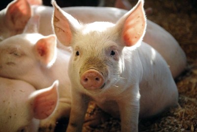
Which Animals and Disease Models Can Be Imaged?
Most preclinical imaging services focus on rodent models (mice and rats), but feasibility extends to larger species depending on instrumentation, available coils or accessories, anesthesia and monitoring protocols, and ethical considerations. Adjustments are made based on the animal’s weight, body temperature regulation, respiratory rate, and fixation requirements during scanning.
Common disease models amenable to imaging include:
Facilities experienced in providing diverse Animal Models In Preclinical Research can advise on which imaging approaches are most appropriate for a given model system, ensuring compatibility between the biological question and the imaging platform.
What Information Should You Prepare About Your Disease Model?
Before approaching an imaging provider, prepare documentation on:
- Expected time points for disease onset, progression, and treatment response
- Clear inclusion and exclusion criteria for enrolling animals into imaging sessions
- Success metrics — what quantitative change constitutes a meaningful biological effect?
Having these parameters defined upfront reduces protocol revisions and accelerates the project timeline considerably.
Ensuring Data Quality and Reproducibility Across Imaging Sessions

Quality and reproducibility in preclinical imaging rely on multiple interrelated factors:
- Standard Operating Procedures for every step of the workflow
- Regular calibration using phantoms
- Standardization of anesthesia and physiological monitoring
- Consistent acquisition parameters
- Clearly defined analysis protocols with appropriate controls
The FDA’s Good Laboratory Practices (GLP) framework provides a regulatory benchmark for nonclinical laboratories, outlining expectations for documentation, traceability, and quality systems.
Practical Measures: Use positive and negative controls within each imaging session, blind analysts to treatment groups where possible, and maintain full documentation of all acquisition parameters. Consistency checks between days, operators, and instruments help identify drift before it compromises data integrity. Providers such as Biotech Farm emphasize well-documented procedures and transparency in collaboration, which directly supports reproducibility across long-term studies.
Which QC Metrics Belong in Every Imaging Report?
Every imaging report should include:
| QC Metric | Purpose |
|---|---|
| Signal-to-Noise Ratio (SNR) | Validates image quality and detection sensitivity |
| Spatial Resolution Verification | Confirms imaging system performance |
| Motion Artifact Assessment | Identifies data that may require exclusion |
| Field Uniformity Metrics | Ensures consistent measurement across image |
| Calibration Deviation Documentation | Tracks any drift from reference standards |
The IAEA’s tutorial resources for PET/CT quality control provide standardized testing frameworks that can be adapted for preclinical scanners.
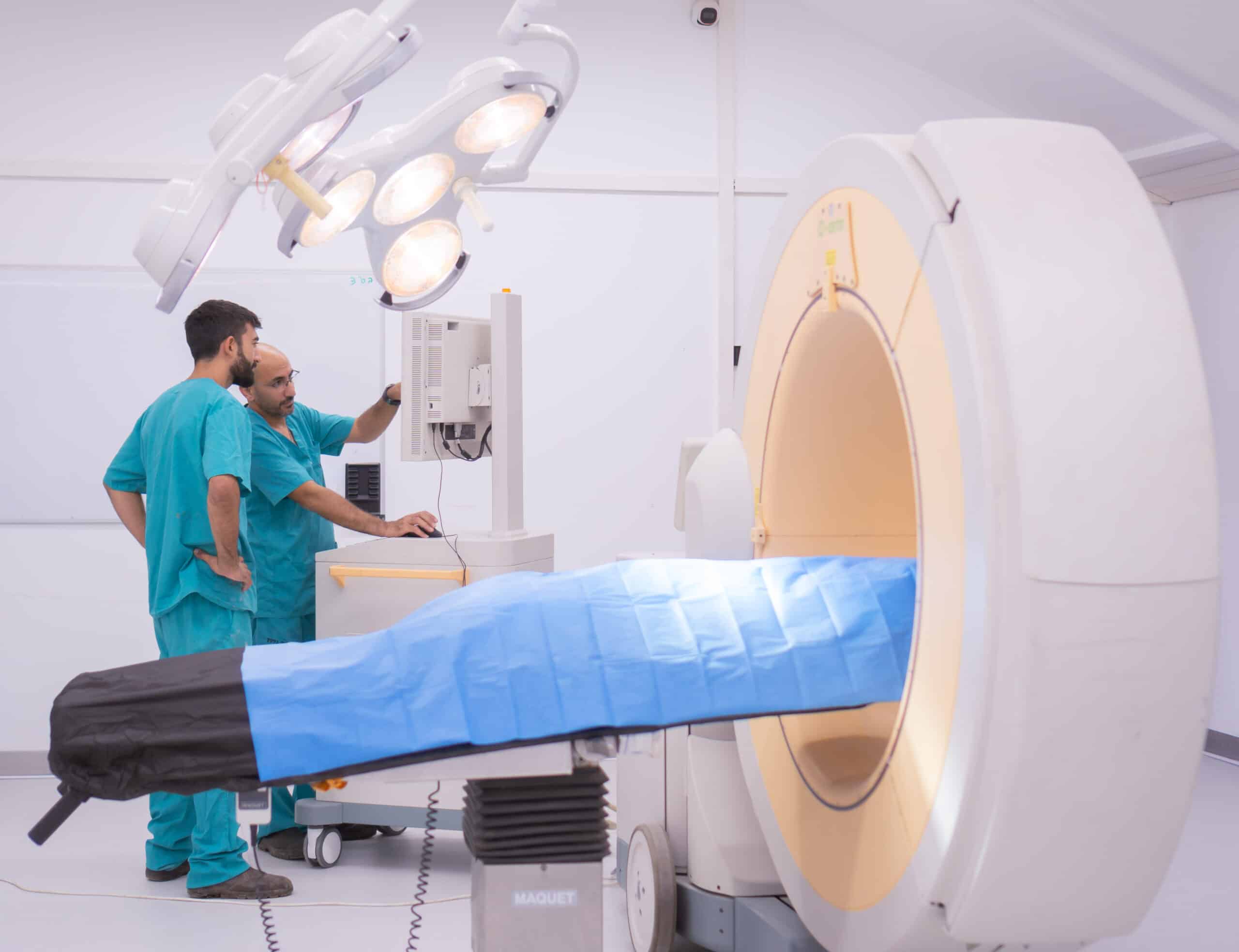
The Four-Phase Workflow: Kickoff Through Final Report
A well-structured imaging project follows a clear four-phase workflow. This end-to-end structure is characteristic of a professional preclinical imaging CRO, where each phase is managed with defined handoffs, quality checkpoints, and documentation standards.
Kickoff Phase
Discuss research question, define quantitative endpoints, establish experimental design, address ethical considerations
Execution Phase
Animal preparation, imaging acquisition according to standardized protocols, real-time physiological monitoring
Analysis Phase
Image processing, quantitative measurement extraction, statistical evaluation with appropriate controls
Reporting Phase
Raw data delivery, processed datasets, detailed methods, interpreted results, regulatory-ready documentation
The ARRIVE guidelines provide a useful framework for ensuring that study plans and reports capture all essential information for transparent and reproducible science.
???? What to Prepare for a Project Kickoff Meeting
Arrive at the kickoff meeting with:
- Clearly articulated biological question
- Preliminary data informing the imaging approach
- Defined budget range and timeline constraints
- Desired primary and secondary endpoints
- Regulatory requirements for the final dataset
Animal Welfare and the 3Rs: How Ethical Compliance Shapes Imaging Study Design
Ethical guidelines and local regulations are strictly followed in all preclinical imaging studies, including approval from the relevant institutional or national ethics body. In Israel, this oversight falls under the Animal Experimentation Council, as detailed in official statistics and regulatory documents published by the Ministry of Health.
The 3Rs framework — Replacement, Reduction, and Refinement — guides every aspect of study design:
???? Replacement
Actively exploring alternatives to animal models wherever scientifically justified
???? Reduction
Optimizing experimental design through longitudinal imaging that allows repeated measurements in the same animal
✨ Refinement
Minimizing pain and distress through appropriate anesthesia, analgesia, and continuous monitoring during scans
Biotech Farm’s commitment to animal welfare and ethical performance, including high standards of care and a transparent 3Rs approach, reflects these principles in daily practice.
What Approvals Are Required Before Any Imaging Can Begin?
All preclinical imaging studies must undergo rigorous review and approval by the local Animal Experimentation Council or equivalent institutional ethics committee (IACUC) before any animal work begins. The approval process evaluates:
- Scientific justification for animal use
- Specific procedures involved in the study
- Measures taken to minimize pain and distress
- Statistical justification for group sizes
Can Imaging Actually Reduce the Number of Animals in Your Study?
Yes. In vivo imaging significantly supports the Reduction principle of the 3Rs by enabling longitudinal studies on the same animal, thereby minimizing the total number of animals required. When each animal serves as its own control — with baseline and post-treatment images compared within the same subject — inter-animal variability is dramatically reduced.
A study that might require 60 animals using terminal endpoint assays at three time points could potentially be conducted with 20 animals imaged longitudinally — a 60%+ reduction in animal use.
— Industry Best Practice Calculation
This aligns with broader regulatory trends, including the FDA’s recent guidance on reducing testing requirements where alternative approaches are scientifically sound.
What Deliverables Should You Expect from a Preclinical Imaging Study?
Deliverables typically include comprehensive documentation that supports both internal decision-making and external regulatory submissions:
| Deliverable Category | What Is Included | Purpose |
|---|---|---|
| Raw Image Data | DICOM files, native scanner formats | Transparency, re-analysis capability |
| Processed Data | Segmented regions, quantitative maps | Direct measurement of endpoints |
| Statistical Analysis | Group comparisons, effect sizes, p-values | Decision-making support |
| Study Report | Full methods, results, interpretation | Regulatory submissions, publications |
| QC Documentation | Calibration records, SNR metrics, artifact logs | Data integrity verification |
A Materials and Methods section written to satisfy transparent reporting standards such as the ARRIVE guidelines 2.0 should be included in every comprehensive study report.
How Should Data Be Presented to Support Go/No-Go Decisions?
Clear visual summaries help stakeholders quickly grasp key findings and their implications for drug development timelines:
- Annotated representative images showing treatment effects
- Quantitative graphs with error bars and statistical annotations
- Concise summary statements for executive review
- Tables comparing treatment groups across time points with highlighted significance
Anesthesia, Monitoring, and Animal Handling During Imaging Sessions
Full-service preclinical imaging providers offer comprehensive animal handling, anesthesia administration, and physiological monitoring throughout every imaging session. This includes:
- Experienced staff for animal preparation, restraint, and post-scan recovery
- State-of-the-art anesthesia delivery systems
- Continuous monitoring of vital signs including body temperature, heart rate, and respiratory rate
As documented in a detailed review in PubMed Central on anesthesia and monitoring during rodent imaging, maintaining stable physiological conditions is essential for generating reliable and reproducible imaging data.
⚠️ Why Consistent Physiological Monitoring Directly Affects Your Data
Variations in body temperature, respiratory rate, or anesthesia depth during a scan can introduce significant artifacts and measurement variability. In longitudinal studies, where comparisons are made across multiple sessions, even small physiological inconsistencies can obscure genuine biological effects. Consistent monitoring protocols ensure that observed changes in imaging metrics reflect true treatment effects rather than physiological noise.
Selecting a Preclinical Imaging CRO in Israel: What Matters Most?
When selecting a preclinical imaging CRO in Israel, evaluate several key dimensions to ensure alignment with your project requirements and quality expectations:
| Evaluation Criterion | What to Look For | Red Flags |
|---|---|---|
| Scientific Expertise | Published studies, experienced team, relevant disease model knowledge | No named scientific leads, no publication record |
| Instrumentation | Well-maintained, calibrated equipment with appropriate accessories | Outdated scanners, no QC documentation |
| Quality Systems | SOPs, calibration logs, GLP-compatible documentation | No written procedures, inconsistent protocols |
| Project Management | Dedicated project manager, clear communication, milestone tracking | No single point of contact, unclear timelines |
| Ethical Compliance | Valid IACUC approvals, documented 3Rs implementation | No ethics documentation, evasive about welfare practices |
| Flexibility | Ability to adapt protocols, accommodate schedule changes | Rigid procedures with no room for scientific discussion |
Biotechfarm: Preclinical Research And Development Services exemplifies the type of facility that combines advanced instrumentation with over three decades of scientific and operational experience in managing complex preclinical programs.
Frequently Asked Questions About Preclinical Imaging Services
Ready to Plan Your Preclinical Imaging Study?
What specific imaging endpoints would advance your current development program, and which modalities are best suited to your biological question? Whether you are designing a first-in-class efficacy study or seeking longitudinal monitoring data for a regulatory submission, having the right preclinical imaging partner is essential for generating actionable, high-quality results.
Reach out to discuss your project requirements and receive a tailored study proposal.