Endoscopic Device Testing in Animals: Preclinical GI Studies That De-Risk Your Path to Market
At the intersection of medical innovation and regulatory rigor, endoscopic device testing in animals represents a pivotal stage in the medical device development lifecycle. Before a novel gastroscope, colonoscope, or therapeutic accessory ever touches a human patient, it must demonstrate safety, performance, and operability under conditions that mimic real clinical use.
Specialized preclinical facilities translate device concepts into validated data packages. With more than 30 years of combined experience in leading large-animal research and a state-of-the-art surgical environment, Biotech Farm supports sponsors who need scientifically robust, well-documented studies to de-risk market entry, accelerate regulatory dialogue, and build investor confidence.
The gastrointestinal tract is a dynamic biological environment with peristalsis, mucus, bleeding tendencies, healing cascades, and immune responses that bench models cannot fully replicate. Preclinical in vivo endoscopic studies are the only credible way to surface hidden design flaws — a sharp edge, an unbalanced tip, an unstable channel — before first-in-human use. Studies conducted with clinical-grade equipment, trained surgical teams, and GLP-aligned documentation produce the regulatory-ready evidence packages sponsors need to advance with confidence.
How is Endoscopic Device Testing in Animals Conducted, Step by Step?
Systematic execution distinguishes a usable dataset from a wasted study. The preclinical pathway for endoscopic devices typically begins with bench testing, advances through simulated and ex vivo models, and culminates in in vivo work in large mammals where procedural translation to humans is critical. Each phase has a defined purpose, and skipping stages often costs more time than it saves.
The process starts by defining intended use and performance requirements, which directly inform model choice and endpoints. Investigators then select between ex vivo tissue platforms and in vivo animal models based on whether the device is being evaluated for material safety, functional performance, or both. Endpoints — safety, efficacy, usability — are pre-specified. The endoscopic procedure is executed by trained operators, and comprehensive data reporting closes the loop, generating documentation that meets regulatory and investor scrutiny.
Step 1: Define Intended Use
Establish performance requirements, clinical claims, and regulatory endpoints that drive model selection and study design.
Step 2: Select the Model
Choose between ex vivo tissue and in vivo large animal based on anatomical fit, lumen diameter, and the specific research question.
Step 3: Execute & Document
Trained operators perform the procedure; all observations, deviations, and measurements are captured in real time for full traceability.
As regulators such as the FDA emphasize, planning, model justification, and full documentation are non-negotiable at every stage of the preclinical pathway for endoscopic devices.
Why Are Animal Studies Necessary for Endoscopic Devices Before Human Trials?
The gastrointestinal tract is not a static tube. It is a dynamic biological environment with peristalsis, mucus, bleeding tendencies, healing cascades, and immune responses that bench models cannot fully replicate. Endoscopic devices interact with this environment continuously, and small design flaws — a sharp edge, an unbalanced tip, an unstable channel — can produce mucosal injury only visible in a living system.
In vivo endoscopic device preclinical studies evaluate mucosal damage, ease of navigation, procedure time, and the device’s ability to perform specific tasks such as resection, ablation, or access. Post-procedure histology then confirms whether the tissue response is acceptable. These studies provide the only credible pre-human evidence of how a device behaves when blood flows, tissue heals, and the operator must react in real time.
“Only a living biological system can reveal the cascade of responses — peristalsis, bleeding, healing, immune activation — that determine whether an endoscopic device is truly safe for human use.”
— Preclinical GI Device Research Principles, Biotech Farm Ltd.
Which Animal Models Are Suitable for GI Device Studies, and Why?
Model selection is rarely about preference; it is about anatomical fit, size compatibility with clinical-grade equipment, and alignment with the research question. A device designed for the human colon must be evaluated in a model whose lumen diameter, wall thickness, and curvature allow meaningful procedural translation.
Porcine models are widely used for colonoscopy device testing because their GI dimensions accommodate human-scale endoscopes, although documented anatomical differences require precise framing of study objectives. Sheep, dogs, and rabbits serve specific niches; rodents support early tissue-level questions.
Large Mammal vs. Small Animal Models: What Each Can Measure
Large Mammals (Porcine, Sheep)
- Human-scale procedural rehearsal
- Compatible with clinical endoscope sizes
- Navigation, energy delivery, survival follow-up
Small Animals (Rodents, Rabbits)
- Early proof-of-concept work
- Mechanism-of-action studies
- Genetic model applications
Ex Vivo, In Vivo, and Survival Studies: What Sets Them Apart?
Sponsors often conflate study types, but each answers a different question. Choosing incorrectly inflates cost without producing usable regulatory evidence. Survival studies are essential when chronic safety or healing must be characterized, since only living follow-up exposes infections, late perforations, or stricture formation.
| Study Type | What It Measures | Key Limitation | Typical Use |
|---|---|---|---|
| Ex Vivo | Material compatibility, immediate tissue effects, basic operability | No circulation, no metabolism, no healing | Early screening, device iteration |
| Acute In Vivo | Real-time procedural performance, immediate tissue response | No long-term healing data | Feasibility, acute safety |
| Survival | Recovery, chronic effects, infection, integration | Longer timeline, higher cost | Pivotal pre-submission studies |
Is GLP Compliance Mandatory for Endoscopic Tool Validation?
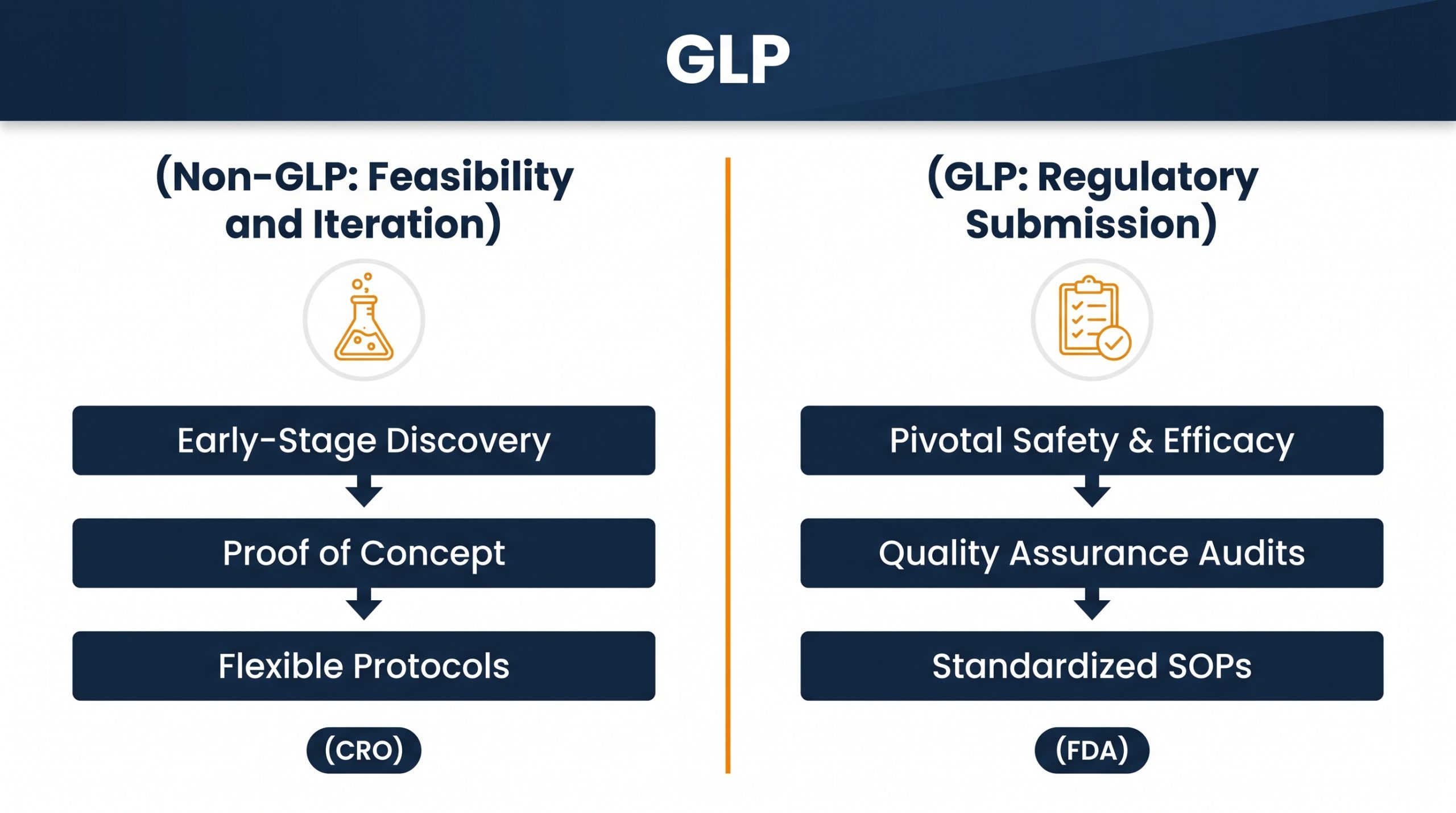
Good Laboratory Practice is not universally required, but it is frequently expected when preclinical data supports a regulatory submission to the FDA, a CE mark dossier, or equivalent. GLP is a quality system covering planning, conduct, monitoring, archiving, and reporting of non-clinical safety studies. Early proof-of-concept work may be conducted non-GLP to preserve flexibility and budget, while pivotal studies feeding regulatory files generally require GLP or GLP-aligned conduct.
Facilities providing services for preclinical GI studies often adhere to GLP principles — a concept crucial for maintaining the credibility of data. This is especially important when considering comprehensive Preclinical Imaging Services for Animal Studies from a Leading CRO.
Choosing GLP vs. Non-GLP by Development Stage
Early Feasibility & Iteration
Flexible, faster, lower cost. Used for proof of concept and protocol refinement. Results are not intended for direct regulatory submission.
Pivotal Regulatory Studies
Required when data feeds an FDA, CE Mark, or equivalent submission. Full QA oversight, archiving, and reporting to internationally recognized standards.
What Endpoints Are Measured in Colonoscopy Device Testing and Similar GI Assessments?
Endpoints translate engineering intent into measurable biology. A well-designed endoscopic study captures safety, performance, and usability in parallel, then ties findings to histology to confirm what the eye and the camera observed. Porcine colonoscopy accessory studies routinely combine procedural metrics with post-procedure histology to confirm that observed performance did not come at the cost of hidden mucosal damage.
| Endpoint Category | Specific Measurements |
|---|---|
| Safety | Mucosal injury, perforation, bleeding severity, adverse events |
| Performance | Procedural efficacy, maneuverability, procedure time, task completion |
| Usability | Operator workload, ease of deployment, intuitive controls |
| Histology | Microscopic damage scoring, healing characterization |
How Long Does a Preclinical Study for an Endoscopic Device Take?
Timelines are driven by study type, device complexity, and regulatory ambition. Ex vivo work can complete in days to a few weeks. Acute in vivo studies typically require several weeks for protocol finalization, ethics approval, execution, and initial reporting. Survival studies extend over weeks to months because animals must be followed through healing, with scheduled imaging, blood work, and sometimes microbiological cultures.
GLP-compliant studies carry additional setup and reporting time due to documentation rigor. Sponsors planning a regulatory submission should budget realistically rather than compress timelines and risk data gaps.
Fast turnaround for material compatibility and basic operability screening.
Includes protocol, ethics approval, execution, and initial data report delivery.
Animal follow-up, serial imaging, histology, GLP documentation, and full study report.
How Many Animals Are Needed for an Endoscopy Device Preclinical Study?
There is no universal number. Sample size flows from the study purpose — pilot, feasibility, or pivotal — combined with expected variability, endpoint type, and required statistical power. Pilot studies often involve small groups to refine the protocol before a larger confirmatory phase.
The 3Rs framework — Replacement, Reduction, Refinement — is embedded in study design. Reduction does not mean using too few animals to draw conclusions; it means using the minimum number that yields scientifically valid, regulator-acceptable data. Justification of the chosen number is itself a documentation requirement that regulators review carefully.
Designing a GI Device Animal Study Protocol for Regulatory and Marketing Data
A protocol that serves both regulators and commercial messaging starts with clear, SMART objectives. The animal model must be scientifically justified against the clinical application. Methodology — anesthesia, procedure steps, device handling, sampling — must be detailed enough for reproducibility. Endpoints and statistical analysis plans are defined up front, not after data is in.
Protocols should reference applicable regulatory guidelines from the outset and incorporate GLP elements when the dataset will support a submission. Marketing claims later in the lifecycle are only defensible if the underlying protocol was tight enough to support them at the time of conduct.
Protocol Design Checklist
- SMART primary and secondary endpoints defined pre-study
- Animal model justification against device intended use
- Statistical analysis plan locked before data collection
- GLP scope decision made at protocol authoring stage
- Reference to applicable FDA/ISO/OECD guidelines included
Ethical Considerations in Endoscopic Device Testing in Animals
Animal research is conducted under strict ethical and legal frameworks. In Israel, the Council for Animal Experimentation at the Ministry of Health governs reporting, approvals, and oversight, with statutory backing through national legislation. IACUC-equivalent committees review every protocol and verify that scientific necessity, animal welfare, and the 3Rs are addressed before any study commences.
Our commitment to ethical practices is integral to our comprehensive Preclinical Research And Development Services, ensuring that all endoscopic device preclinical studies are conducted with the highest standards of animal welfare, transparent documentation, and accountable oversight.
Balancing Innovation with Animal Welfare
Advancing medical technology and protecting animal welfare are not opposing goals. Loving tender care of the animals, well-documented procedures, and transparent collaboration with sponsors are operational expressions of the same ethical standard. Every protocol undergoes IACUC-equivalent review, and humane endpoints are defined in advance — not as an afterthought.
What Role Does Imaging Play in Endoscopic Device Testing?
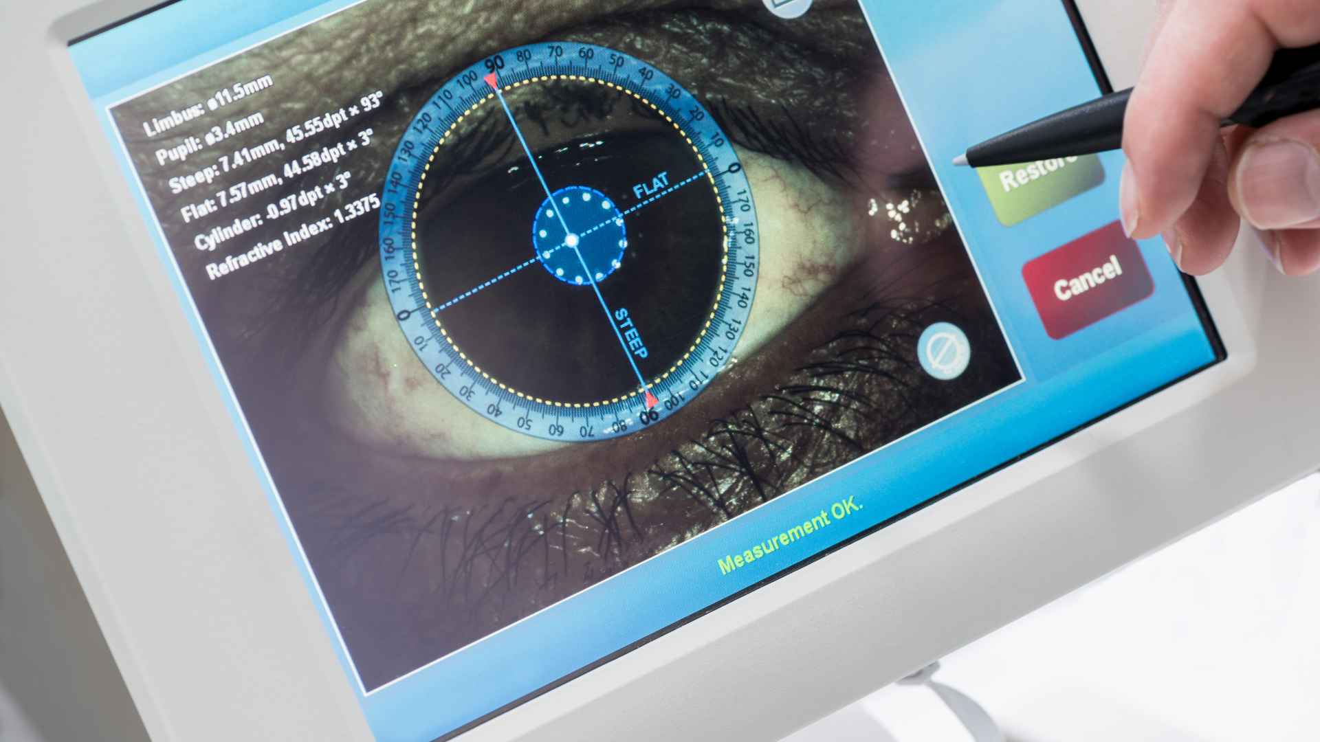
Imaging transforms procedural observations into objective, quantifiable data. Intra-procedural fluoroscopy and high-definition endoscopy track device deployment, navigation, and interaction in real time. Post-procedural CT, MRI, and ultrasound document tissue response, healing trajectories, and any structural complications.
For endoscopic tool validation, imaging complements visual assessment and histology, producing a multi-modal data package that withstands regulatory scrutiny. Integrated imaging is particularly valuable in survival studies, where serial scans replace repeated invasive sampling.
C-Arm fluoroscopy, 4K HD endoscopy — real-time tracking of device deployment and navigation.
CT, MRI, high-definition ultrasound — tissue response, healing trajectories, and structural complications.
Microscopic tissue analysis confirming macroscopic findings and characterizing healing at the cellular level.
Common Mistakes Sponsors Make in Preclinical Endoscopic Studies
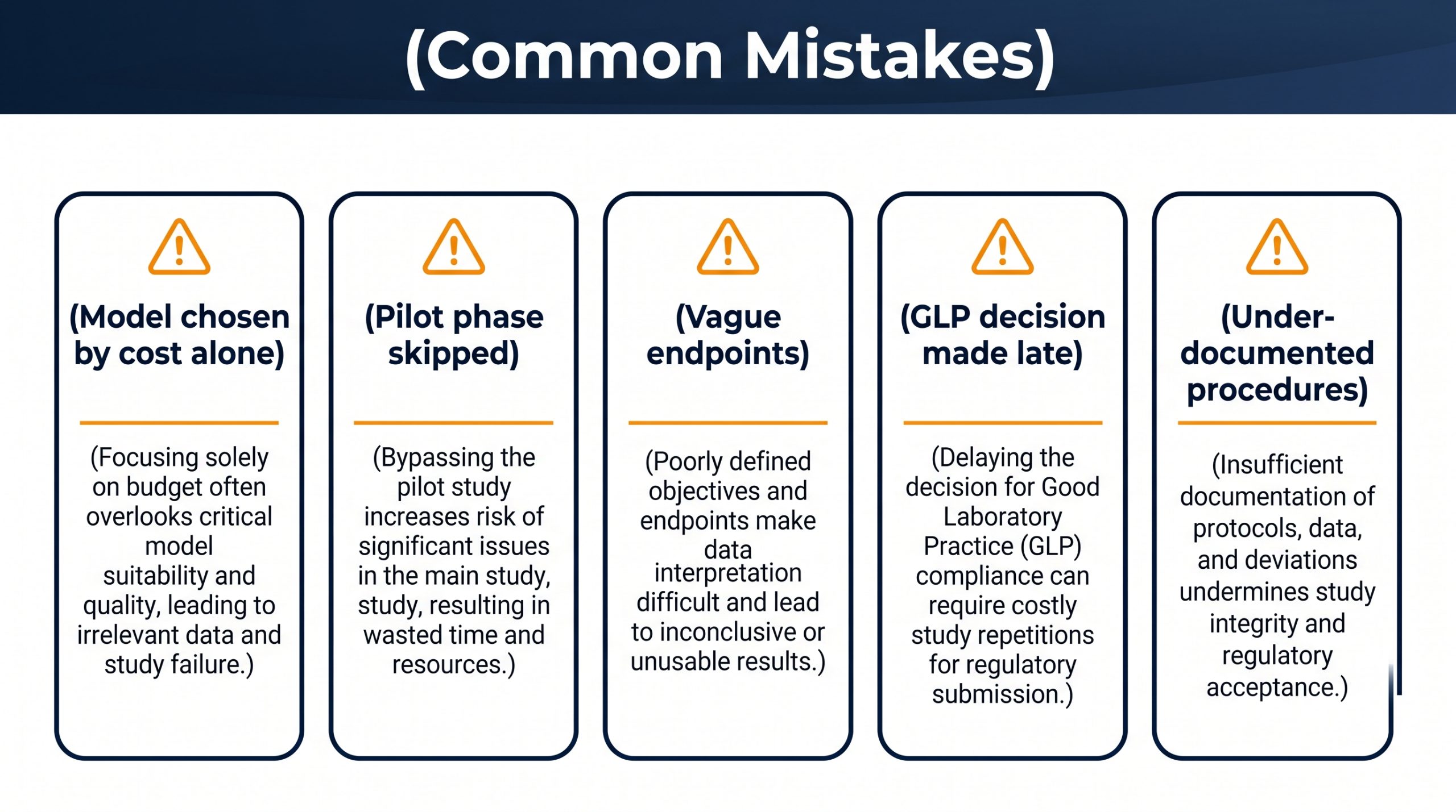
Several recurring errors inflate cost and weaken regulatory positioning. Recognizing them early protects the program and preserves the integrity of the evidence package.
| Common Mistake | Consequence | Preferred Practice |
|---|---|---|
| Choosing model based on cost alone | Anatomical mismatch invalidates findings | Justify model against device intended use |
| Skipping pilot before pivotal | Protocol flaws surface in GLP study | Run a small feasibility phase first |
| Vague endpoints | Data cannot support claims | Pre-specify SMART endpoints |
| Late GLP decision | Re-doing the study under GLP | Decide GLP scope at protocol design |
| Under-documented procedures | Regulator queries and delays | Capture raw data and deviations live |
Sponsors planning a regulatory submission should budget realistically rather than compress timelines and risk data gaps. A study that requires repetition due to documentation failures costs multiples of the time saved by skipping proper setup phases.
Translating Animal Study Results to Human Applications
Translation depends on understanding what the model can and cannot represent. Comparative anatomy and physiology dictate which findings extrapolate cleanly and which require caveats. Porcine GI work, for example, supports strong feasibility and safety conclusions for human colonoscopic systems while acknowledging documented anatomical differences.
Animal data primarily delivers proof of concept and early safety signals. It informs risk assessment for first-in-human trials, guides design iteration, and surfaces issues that would otherwise emerge in clinical use. Iterative testing — design, evaluate, refine, retest — remains the safest route from prototype to patient.
“Iterative testing — design, evaluate, refine, retest — remains the safest and most cost-effective route from endoscopic device prototype to first-in-human clinical use.”
— Adir Koreh, CEO, Biotech Farm Ltd.
Mapping Sponsor Needs to On-Site Capabilities
A practical way to evaluate a preclinical partner is to map your needs against what the facility delivers in practice. The table below reflects Biotech Farm’s operational capabilities.
| Sponsor Need | How a Specialized Facility Delivers |
|---|---|
| Human-scale procedural rehearsal | Large animal models and clinical-grade endoscopy suites |
| Multi-modal data capture | Fluoroscopy, ultrasound, endoscopic HD video, histology |
| Regulatory-ready documentation | Well-documented procedures aligned with GLP principles |
| Ethical oversight | Animal welfare protocols, IACUC-equivalent review, 3Rs application |
| Scientific dialogue | Interactive conference space and scientific escort throughout the study |
| Fit for sponsors operating in Israel | Local regulatory familiarity and senior surgical crew on site |
Documentation Required for Regulatory Submission of Preclinical Data
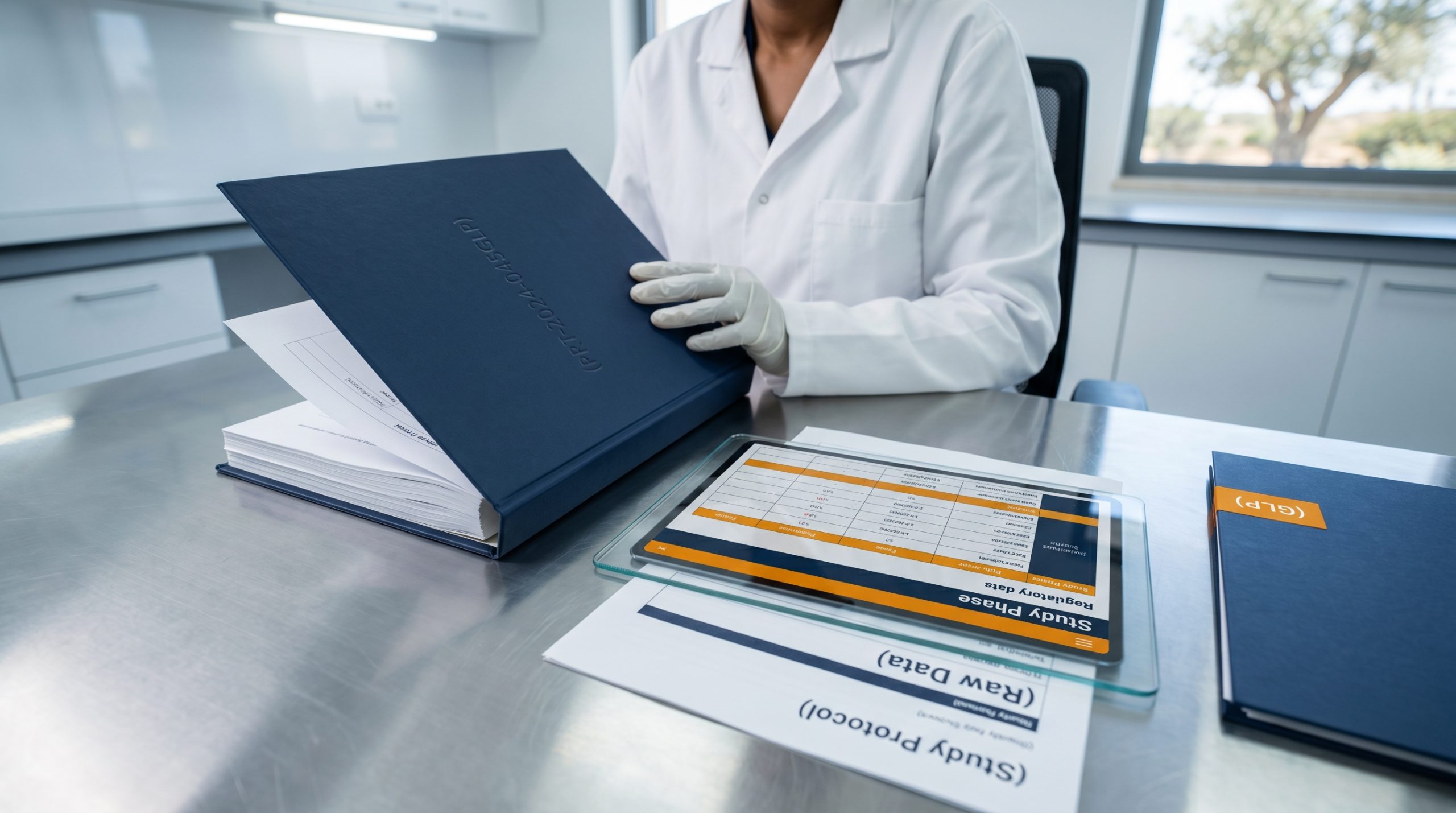
Regulators expect a complete, traceable evidence chain. The study protocol defines design, objectives, and endpoints. Raw data captures every observation and measurement. The study report integrates methodology, results, discussion, and conclusions — including findings specific to colonoscopy device testing where relevant. Ethical approvals and, where applicable, GLP compliance records complete the package.
Design, objectives, endpoints, model justification, statistical analysis plan — all locked before study start.
All observations, measurements, and protocol deviations captured live with timestamps for full traceability.
IACUC-equivalent approvals, GLP facility accreditation, personnel qualifications, and QA audit trail.
Key principle: Missing or inconsistent documentation is a leading cause of regulatory delay. Capturing data correctly during conduct is far cheaper than reconstructing it afterward. A complete, traceable evidence chain is not a bureaucratic exercise — it is the foundation on which regulatory confidence is built.
What Are the Main Challenges in Endoscopic Device Animal Studies?
Sponsors face model limitations, ethical responsibilities, cost pressures, evolving regulatory expectations, and a need for specialized expertise. No model perfectly mimics human physiology, and acknowledging that limitation is itself a sign of methodological maturity. Cost and time scale with study complexity, and the regulatory landscape shifts across markets.
Challenges include the need for specialized equipment such as a professional colonoscope and highly trained teams for both procedural aspects and ethical oversight of endoscopic tool validation. Sponsors who address these requirements early avoid late-stage rework.
No animal model perfectly replicates human GI physiology. Transparent study framing is essential.
FDA, CE Mark, and international requirements differ. Protocol design must account for the target submission market.
Clinical-grade colonoscopes, fluoroscopy, and 4K imaging require a fully equipped large-animal facility.
Why Partner with a Specialized CRO for Endoscopic Device Testing in Animals?
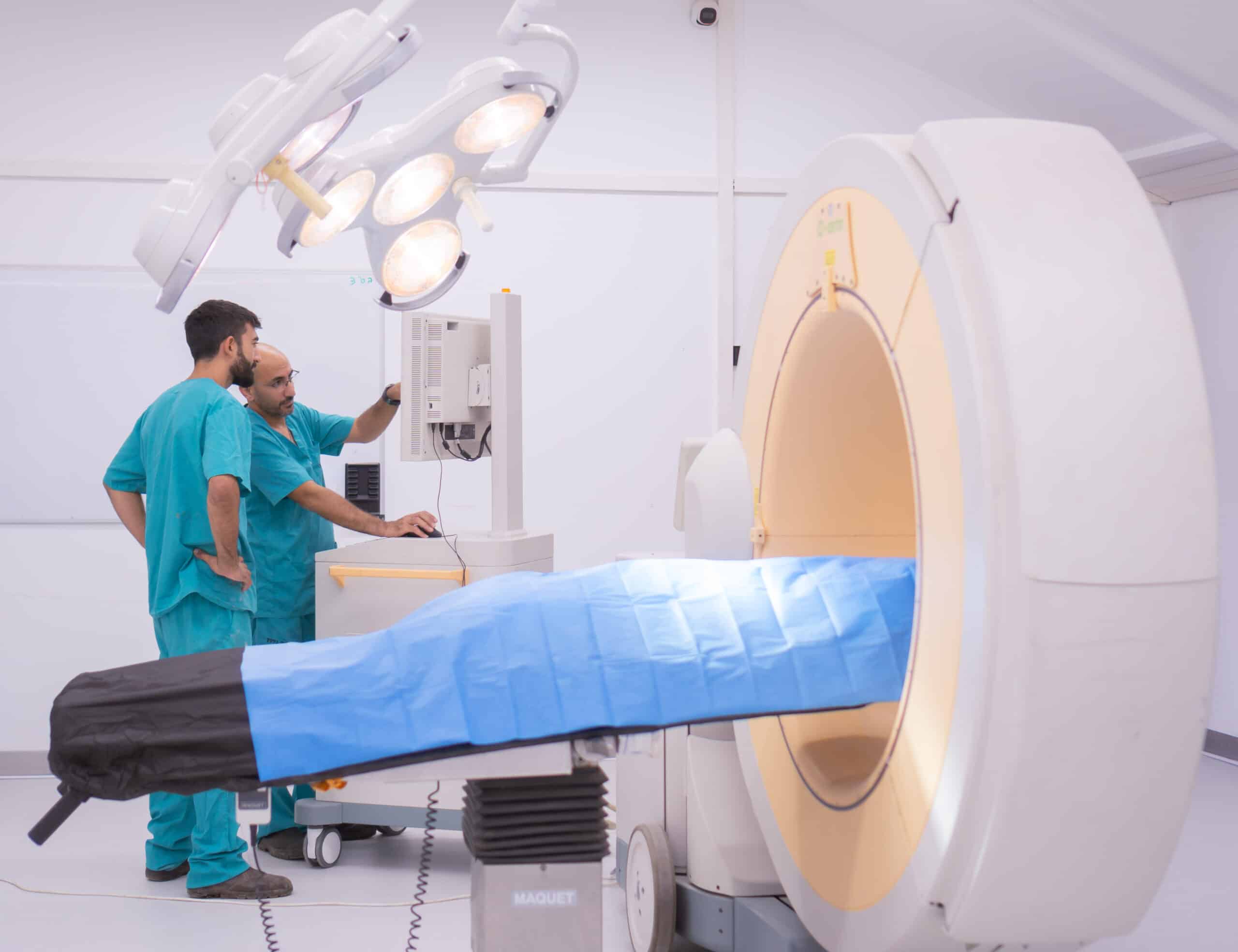
A specialized CRO concentrates the rare combination of scientific expertise, surgical capability, regulatory literacy, and animal welfare governance that endoscopic device studies require. Sponsors gain access to senior surgeons, veterinary teams experienced in GI device animal study work, and facilities equipped with C-Arm fluoroscopy, high-definition ultrasound, and 4K laparoscopic towers.
Beyond infrastructure, an experienced partner contributes a tailored approach — matching needs to services — that compresses timelines and reduces avoidable iterations. Studies aligned with international protocols, such as endoscopic device investigations registered on public clinical trial databases, illustrate how rigorously designed preclinical work feeds clinical evaluation. CROs are also instrumental in bringing advanced endoscopic technologies through preclinical phases efficiently while maintaining transparency in collaboration.
- 20+ years senior large-animal surgical expertise
- C-Arm fluoroscopy, HD ultrasound, 4K laparoscopic imaging on site
- GLP-aligned documentation and full regulatory package delivery
- IACUC-equivalent ethics oversight with embedded 3Rs methodology
- Interactive sponsor collaboration space for live scientific dialogue
Frequently Asked Questions
Ready to De-Risk Your Endoscopic Device Pathway?
Where is your device in its preclinical journey, and what evidence will your next regulatory conversation demand? If you are planning endoscopic device testing in animals and want a scientifically supportive partner with a fully equipped large animal facility, senior surgical expertise, and a documented commitment to animal welfare — let’s connect.