Preclinical Large-Animal Research · Regulatory-Grade Evidence
Pig Model Wound Healing: Preclinical Skin Studies That Actually Translate to the Clinic
Built on rigorous protocol design, quantitative histology and more than three decades of hands-on large-animal research experience. Led by Adir Koreh, CEO of Biotech Farm Ltd. and owner of Biotech Anatomy Ltd., with 20+ years directing large-animal model set-up alongside a veterinary team that has worked together for more than a decade.
Wound care products live or die on evidence. A dressing, a hydrogel, a growth factor, a cell therapy or an antimicrobial coating may perform beautifully in a dish and still fail once it meets a living, vascularized, immunologically active tissue. The pig model wound healing platform sits exactly at that junction: close enough to human skin to be predictive, controlled enough to be measurable, and robust enough to support regulatory submissions. This guide walks through what a porcine wound model is, how wound types differ, which endpoints matter beyond simple closure, and how to structure a study that produces data your reviewers, investors and notified body will accept.
Exclusive Insight
Comparative work has reported substantially higher concordance between porcine and human wound healing outcomes than between small mammals and humans — a finding that has quietly reshaped preclinical wound science for two decades. The practical takeaway: effect sizes measured in a well-run pig skin wound healing study tend to survive translation to the clinic, which reduces the risk of building an entire clinical program on a biological artifact.
What Is Pig Model Wound Healing?
Pig model wound healing is a preclinical in vivo approach in which standardized, controlled wounds are created on porcine skin and then monitored over time to quantify both the rate and the quality of repair. The wounds are treated with the investigational device, dressing, biomaterial or active ingredient, compared against untreated or standard-of-care controls, and evaluated using imaging, planimetry and histology.
The reason this model earns its place in development programs is mechanistic. Porcine skin closes partial-thickness defects mainly through re-epithelialization, much like human skin, rather than through the aggressive contraction seen in loose-skinned rodents. That single difference changes how a product’s effect is expressed, measured and interpreted, and it is why a swine dermal wound study is often the last preclinical step before first-in-human work.
Why Do Pigs Outperform Rodents in Skin Wound Healing Studies?
Comparative work has reported substantially higher concordance between porcine and human wound healing outcomes than between small mammals and humans, a finding that has shaped the field for two decades (Sullivan et al., Wound Repair and Regeneration). The similarities are structural as well as functional: comparable epidermal and dermal thickness, sparse hair coat, similar dermal collagen biochemistry, comparable vascular organization of the follicular unit, and the same overlapping healing phases.
For a sponsor, this translates into a practical benefit. Effect sizes measured in pig skin wound healing studies tend to survive the jump to clinical evaluation, which reduces the risk of building a clinical program on a biological artifact.
| Feature | Small mammals | Pig | Human |
|---|---|---|---|
| Epidermal thickness | Very thin, few cell layers | Comparable to human | Reference |
| Dominant closure mechanism | Contraction of loose skin | Re-epithelialization | Re-epithelialization |
| Dermal adherence | Loose, mobile panniculus carnosus | Firmly attached | Firmly attached |
| Wound size feasible | Small, limited replicates | Multiple clinically sized wounds | Clinical dimensions |
| Translational concordance | Lower | Higher | — |
Which Wound Model Fits Your Product: Excisional, Incisional or Burn?
There is no universal wound. The model should be selected from the claim you intend to support, not from convenience. An epithelializing dressing needs a tissue-deficient defect; a surgical sealant needs an apposed incision; an antimicrobial needs a wound bed that can genuinely become colonized. Choosing correctly at the protocol stage prevents the most expensive failure in preclinical work, which is a technically flawless study that answers the wrong question.
| Model | Primary research question | Typical endpoints |
|---|---|---|
| Partial/full-thickness excisional | Secondary intention healing, epithelial coverage | Wound area, % re-epithelialization, neodermis thickness |
| Incisional | Primary healing, scar formation, mechanical integrity | Tensile strength, scar width, dehiscence rate |
| Thermal burn | Depth progression, debridement, infection control | Burn depth conversion, bioburden, graft take |
When an Excisional Model Is the Right Choice
Excisional wounds remove tissue and force the body to rebuild it. Partial-thickness excisions created with a dermatome remove the epidermis and a defined portion of dermis while sparing follicular and glandular remnants, closely mimicking split-thickness graft donor sites. Full-thickness excisions extend through the dermis and are preferred for scaffolds, dermal substitutes and regenerative matrices where neodermis formation, angiogenesis and remodeling are the real story.
When an Incisional Model Is the Right Choice
Incisional wounds are the model of choice when the product interacts with apposed tissue edges: sutures, staples, adhesives, sealants and anti-scar formulations. Here the informative endpoints are biomechanical and structural — tensiometry, scar width and collagen fiber orientation — rather than planimetric. Paired designs, in which treated and control incisions sit on the same animal along matched anatomical lines, sharply reduce inter-animal variability.
When a Burn Model Is the Right Choice
Burn models reproduce thermal injury with a controlled contact device delivering defined temperature, pressure and dwell time. They carry devitalized tissue, a persistent inflammatory drive and high susceptibility to colonization. If your claim concerns infected or complicated wounds, a clean excisional bed will flatter your product and mislead your clinical planning.
Porcine Wound Model Versus Swine Dermal Wound Study: A Distinction Worth Keeping
The two terms are often used interchangeably, but they describe different objects. A porcine wound model is the biological and technical construct: species, strain, anatomical site, wound geometry and depth, injury method and standard care. A swine dermal wound study is the scientific investigation built on top of that construct: hypothesis, randomization, allocation, sampling schedule, analytical methods and statistical plan.
Why does the distinction matter? Because model validation and study validity are audited separately. A poorly characterized model undermines even an elegantly designed study, while a validated model can still yield uninterpretable data if the study design is weak.
Designing a Pig Skin Wound Healing Study for Regulatory and Commercial Relevance
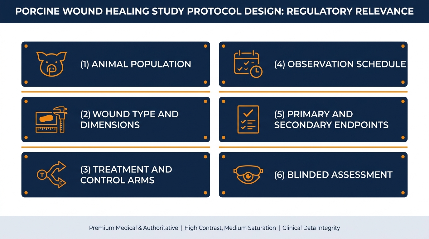
A defensible design begins by writing the intended claim as a sentence, then working backwards to the measurement that supports it. From there you define the animal population, wound type and dimensions, anatomical map, treatment and control arms, dressing regimen, observation schedule, and primary and secondary endpoints. Standardized porcine protocols emphasize consistent wound siting, uniform depth and repeatable sampling and histology as the three levers that most reduce variance (standardized partial-thickness porcine model, IJMS).
Protocol Parameters You Should Never Leave Undefined
Methodology checklist: wound diameter and depth; inter-wound spacing; dorsal versus flank placement and randomization of positions; anesthesia and analgesia regimen; dressing type, change frequency and fixation method; time points for photography and biopsy; biopsy orientation and embedding plane; stains and morphometric definitions; predefined exclusion criteria. Animal welfare and the 3R principles belong in the same document, not in an appendix.
How Is Wound Closure Measured in a Pig Model?
Closure is measured on two levels that must agree before you report a result. The macroscopic level uses standardized digital photography with a scale reference, followed by planimetry to calculate wound area at each time point, normalized to day-zero area. The tissue level uses histology to determine whether a confluent neo-epidermis actually bridges the defect, since a wound can look closed clinically while remaining incompletely epithelialized microscopically.
In a well-run pig wound closure model, camera distance, lighting, lens and marker placement are fixed by standard operating procedure, and images are analyzed by an assessor blinded to treatment allocation. Without those controls, the variance introduced by photography alone can exceed the treatment effect you are trying to detect.
Area or Width: Two Ways to Report the Same Wound
Planimetric area is intuitive, non-invasive and allows repeated measurement of the same wound across the entire study. Its weakness is that it is a surface projection and cannot distinguish true epithelial coverage from contracted margins or fibrin cover. Histological width measurement in cross-section reports the linear distance covered by neo-epidermis relative to the original defect edge — precise and mechanistically meaningful, but terminal and section-dependent. Modern practice combines both: planimetry for kinetics, morphometry at defined sacrifice points for confirmation.
What Exactly Is Re-epithelialization, and How Is It Quantified?
Re-epithelialization is the migration and proliferation of keratinocytes from wound margins and surviving adnexal structures until a continuous epidermal sheet covers the defect and a basement membrane is re-established. It is commonly reported as the percentage of the original wound width covered by neo-epidermis in representative histological sections, sometimes accompanied by neo-epidermal thickness and cell-layer counts.
Beyond Closure: The Endpoints That Demonstrate Regeneration Quality
Faster is not automatically better. A wound that closes rapidly with a thin, disorganized dermis and a hypertrophic scar may be commercially weaker than one that closes two days later with a thick, well-vascularized neodermis. Endpoints that capture quality include neodermis thickness, granulation tissue area and maturity, vessel density, inflammatory infiltrate composition, collagen organization under polarized light, myofibroblast presence, and biomechanical strength in incisional or grafted designs.
- Neodermis thickness and vascularization
- Collagen organization under polarized light
- Inflammatory infiltrate composition
- Biomechanical tensile strength (incisional models)
How Long Does a Porcine Wound Study Take?
Timelines follow biology, not calendars. Superficial and partial-thickness excisional wounds typically reach substantial re-epithelialization within roughly one to two weeks, so studies commonly use time points at days 3, 7, 10 and 14. Full-thickness defects and burns require longer observation, often three to six weeks, to capture granulation, contraction and early remodeling. Scar quality and matrix maturation may justify extending to two or three months. Add acclimatization, surgical scheduling, histology processing and reporting, and a realistic end-to-end program usually spans several months from protocol approval to final report.
How Many Wounds per Animal Before Data Quality Suffers?
Multiple wounds per animal are standard practice and are one of the main reasons the porcine platform is statistically efficient. Optimized wound array designs in miniature pigs have demonstrated that dozens of standardized wounds can be created and evaluated on a single animal when spacing, symmetry and site randomization are controlled (wound array model, Scientific Reports).
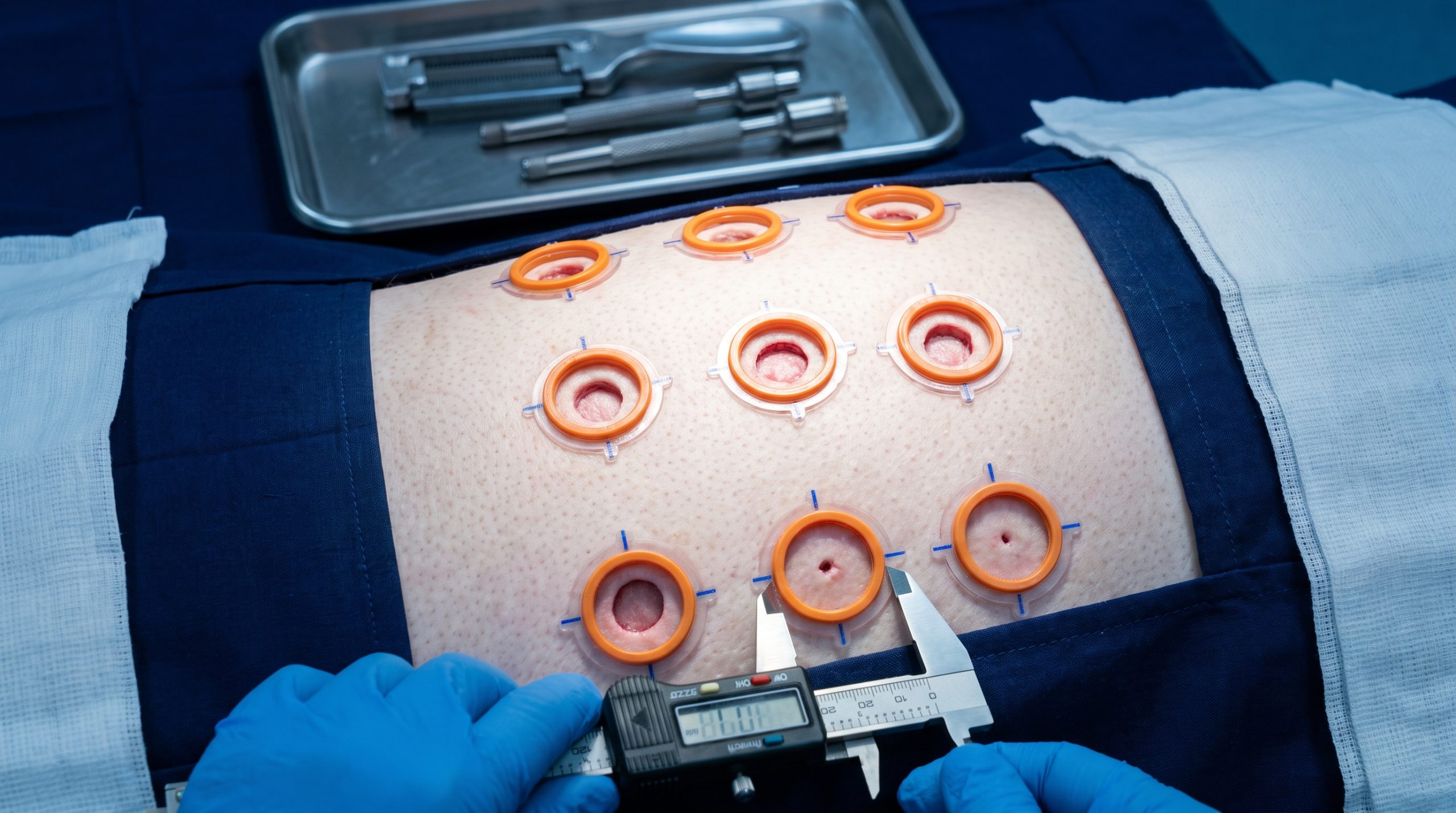
The constraints are real, though. Wounds placed too closely can influence each other through shared inflammatory and vascular fields. Anatomical position affects healing rate, so treatments must be rotated across positions rather than assigned to a convenient region. Dressing footprint also limits density, since overlapping dressings compromise fixation and cross-contaminate treatments.
The 3R Angle: Fewer Animals, Same Statistical Power
Placing several wounds on each animal is not only an efficiency measure; it is a Reduction strategy under the 3R framework. Within-animal designs remove a large share of biological variability, which means the same statistical power can be achieved with fewer animals. Refinement follows through positive-reinforcement handling, appropriate multimodal analgesia, well-tolerated dressing regimens and clearly defined humane endpoints.
At our facility, welfare is treated as a scientific requirement rather than a compliance formality. Calm, well-socialized animals move less under dressings, retain them longer and generate cleaner data. Welfare practice and data quality point in the same direction.
Minipig or Domestic Pig: Which Animal Should You Choose?
Minipig strains such as Göttingen and Yucatan offer genetic uniformity, predictable adult size, easier long-term housing and simpler handling, which makes them attractive for studies running beyond a few weeks or requiring frequent manipulation. Domestic pigs offer a larger available skin surface, supporting more wounds per animal or clinically sized defects, and they are often more readily available and less costly to source. Their rapid growth is the main caveat: over a long study, body wall expansion can distort wound geometry. The decision should be made before the protocol is drafted, driven by study duration, required wound number and dimensions, logistics and budget.
Phase One: Screening
Six formulations tested on porcine skin explants for antimicrobial activity and tissue compatibility — three eliminated.
Phase Two: Exploratory In Vivo
Standardized partial-thickness excisional wounds compared across days 3, 7, 10, 14. One candidate shows a consistent two-day acceleration.
Phase Three: GLP Pivotal
Expanded group sizes, blinded pathology, full morphometry and safety assessment under GLP conditions.
Common Mistakes That Quietly Ruin Porcine Wound Studies
The most frequent failures are mundane. Dressings that migrate or are removed by the animal, producing missing data at the exact time point that mattered. Uncontrolled depth, when the dermatome or biopsy punch is not calibrated on each animal. Unblinded assessment of photographs. Biopsies taken off-axis, so the epithelial tongue is measured obliquely. Undocumented infection treated as background noise. And, historically, the absence of a shared standard, which has long made cross-study comparison difficult.
Practical Controls That Preserve Data Integrity
Validate wound creation on a pilot animal and confirm depth histologically. Fix dressings with a documented, tested layering system and inspect at defined intervals. Randomize treatment to anatomical position and include an internal control wound on every animal. Blind the image analyst and pathologist. Record every deviation in the raw data. Predefine exclusion criteria before the study starts.
What a GLP Wound Healing Study in Pigs Usually Includes
When data are intended for a regulatory dossier, the study is conducted under Good Laboratory Practice, an internationally harmonized quality framework covering organization, planning, performance, monitoring, recording, reporting and archiving of non-clinical safety studies (OECD Principles on Good Laboratory Practice). GLP does not make a study scientifically better on its own; it makes the results traceable, reconstructable and auditable.
In practice, sponsors often run an exploratory non-GLP study first to establish dose, timing and feasibility, then repeat the pivotal design under GLP conditions.
“A named study director, an independent Quality Assurance Unit, approved SOPs and contemporaneous raw data are what turn a good experiment into evidence a reviewer can trust.” — OECD Principles on Good Laboratory Practice
Ex Vivo Porcine Skin Versus In Vivo: What Does Each Actually Prove?
Ex vivo porcine skin explants are excellent screening tools. They are inexpensive, high throughput and highly controlled, and they support permeation studies, short-term cytotoxicity assessment, antimicrobial and anti-biofilm testing, and mechanism-of-action work on isolated tissue. What explants cannot provide is circulation, systemic immunity, innervation, angiogenesis or true remodeling.
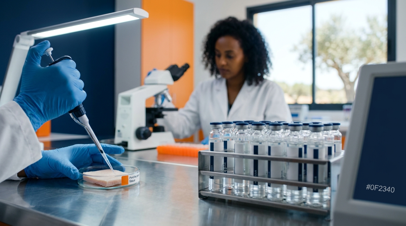
Any claim that depends on the host response requires an in vivo porcine wound model. The efficient strategy is sequential: use ex vivo work to narrow candidates and define concentrations, then commit in vivo resources to the two or three formulations that survived screening.
How Biotech Farm Supports Porcine Wound Healing Programs
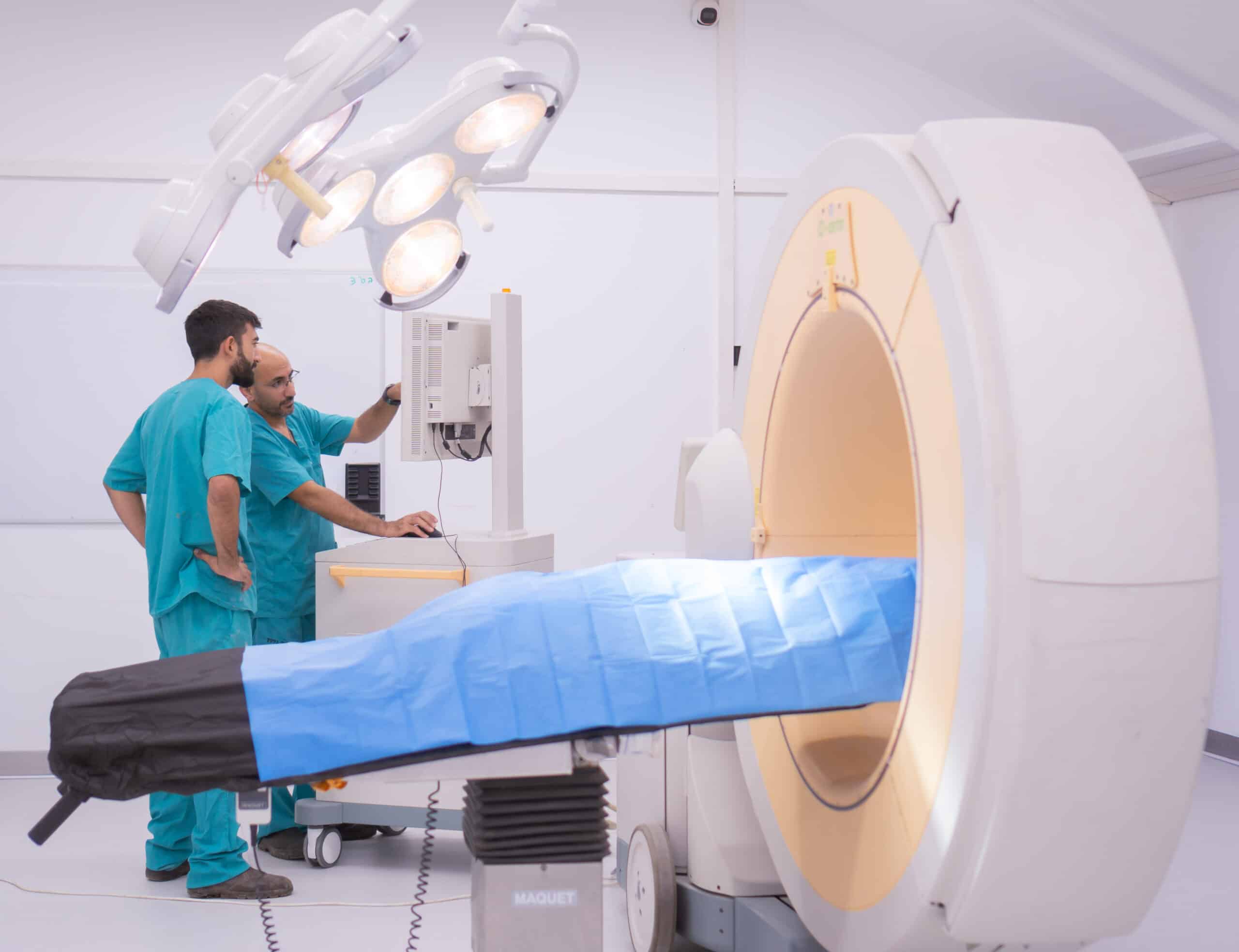
Biotech Farm is a large-animal preclinical facility built around scientific partnership rather than task execution. Programs are designed together with the sponsor, the study team provides scientific escort through protocol drafting, ethics submission, in-life conduct and reporting, and the facility infrastructure supports surgical precision and imaging on site.
| Development need | How the facility supports it |
|---|---|
| Choosing the right model | Joint protocol design matching wound type, depth and endpoints to the indication |
| Reproducible wound creation | Fully equipped surgery rooms, calibrated instrumentation, experienced surgical crew |
| Evidence of regeneration quality | Planimetry, standardized photography, quantitative histology and morphometry |
| Regulatory readiness | Documented procedures and study structures aligned with regulatory expectations |
| Ethical and welfare compliance | 3R-oriented designs, spacious housing, attentive veterinary care |
| Program agility | Flexible, tailored formats for exploratory, pivotal and comparative work |
Comfortable animal housing, gentle daily handling and a team that knows each animal reduce stress responses, improve dressing retention and produce more consistent healing curves. The dermatological platform sits alongside cardiology, orthopedics, ophthalmology and metabolic research capabilities. Further detail is available on the Wound Healing and Dermatological Models page.
Frequently Asked Questions
Which Question Is Your Next Study Actually Meant to Answer?
If you can articulate that in one sentence, the model, the endpoints and the timeline follow logically from it. If you cannot yet, that is precisely the conversation worth having before any animal is enrolled. The team at Biotech Farm works with sponsors to translate a product concept into a defensible pig model wound healing protocol, execute it in a fully equipped large-animal facility with experienced surgeons and attentive veterinary care, and deliver quantitative, well-documented data that stands up to scientific and regulatory review.