Pig Model Orthopedic Device Testing: A Practical Guide to Porcine Bone Studies
Preclinical planning, endpoints, and regulatory expectations for orthopedic implants
Orthopedic device development stands or falls on the quality of the preclinical evidence behind it, and that evidence depends on scientific judgment accumulated over years of hands-on surgical and study-management experience. A porcine study is never only a surgery; it is a chain of decisions — model selection, defect geometry, fixation strategy, imaging intervals, histology processing, biomechanical endpoints and documentation — where each link determines whether reviewers can rely on the result. Teams that combine senior surgical hands, veterinary care, and regulatory literacy tend to produce data that translates. This guide walks through the practical anatomy of pig model orthopedic device testing, from choosing the species and the bone site to reporting the study in a form that supports a medical device submission, with attention to animal well-being and transparent, well-documented procedures throughout.
Exclusive Insight: The Convergence Rule
No single modality proves osseointegration on its own. Reviewers look for convergence — micro-CT bone volume, histomorphometric bone-implant contact, and mechanical pull-out or removal torque all pointing the same direction at the same timepoint. Studies that pre-specify this triad before surgery, rather than reconstructing it afterward, consistently generate fewer regulatory information requests.
Why choose a pig model for orthopedic device testing?
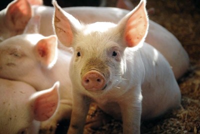
Pigs are used as a translational bridge because their skeletal anatomy, bone remodeling activity and body mass fall within a range that is meaningful for human orthopedics. Pig model orthopedic device testing allows a device to be implanted at close to its clinical dimensions, loaded by a comparable body weight, and evaluated for bone-implant interaction under physiologically relevant conditions. Reviews of animal models in preclinical orthopedic implant research consistently point to large animals when the question involves load transfer, cortical healing and long-term integration rather than isolated cellular response. For orthopedic implants such as plates, intramedullary devices, screws, spinal cages and bone substitutes, a large preclinical model supports simultaneous assessment of surgical handling, biomechanics and healing biology in a single, well-controlled study.
What makes the porcine bone model ideal for orthopedic research?
The porcine bone model offers cortical thickness, trabecular architecture, mineral density and lamellar organization that resemble human bone more closely than smaller laboratory species. Pigs also remodel bone briskly, which is why swine bone regeneration data can be generated within study windows that remain practical for a development program. Rapid somatic growth is both an advantage and a constraint: it shortens timelines but must be controlled through breed and age selection so that skeletal maturity does not drift during the follow-up period. Availability, established husbandry, predictable temperament under trained handling, and well-characterized surgical anatomy make the species a practical choice ethically and operationally. For a pig fracture healing model, these traits translate into reproducible callus formation, measurable bridging and consistent inter-animal comparability when handling, nutrition and analgesia are standardized.
Pig vs. sheep model for orthopedic implants: which fits your study?
The pig vs sheep model for orthopedic implants question rarely has a universal answer; it depends on the endpoint that drives your submission. Pigs are often selected when bone dimensions, marrow cavity size and active remodeling are decisive, which is central to quantifying swine bone regeneration and early osseointegration. Sheep are frequently used for long-bone weight-bearing models, ovine spine work and long-duration follow-up where slower, more static remodeling is desirable. Practical factors — housing, containment of implanted limbs, post-operative mobility, and the feasibility of repeated imaging — also weigh heavily.
| Decision factor | Porcine model tendency | Ovine model tendency |
|---|---|---|
| Bone remodeling rate | Fast; suits early integration questions | Slower; suits long-term stability |
| Growth during study | Rapid; requires age/breed control | More stable in adults |
| Cortical architecture | Close to human lamellar bone | Denser, more plexiform in areas |
| Weight-bearing long-bone models | Feasible with careful fixation | Widely established |
| Handling and repeated imaging | Trainable, sedation-friendly | Manageable, calmer restraint |
In vivo or ex vivo: two complementary testing routes
In vivo testing means the device is implanted in a living animal and evaluated over time, capturing osseointegration, inflammatory response, callus maturation, infection risk and functional loading. Ex vivo testing uses harvested bones or limb segments to characterize mechanical behavior without a biological time course; published work on ex vivo biomechanical bone testing of pig femur illustrates how three-point bending and comparable setups quantify stiffness and failure loads. Ex vivo work is efficient for screening instrumentation, validating insertion torque, refining surgical technique and rehearsing fixation before any animal is enrolled. A pig fracture healing model, by contrast, requires in vivo follow-up because the outcome of interest is biological bridging over weeks. Most robust programs sequence both: ex vivo to de-risk, in vivo to demonstrate.
Designing a successful pig fracture healing model
Model design begins with the clinical scenario you intend to mimic. A transverse osteotomy with rigid plate fixation answers different questions than a comminuted metaphyseal injury or a segmental gap. Key design decisions include defect geometry and reproducibility, use of jigs or saw guides, fixation stiffness, soft-tissue handling, and whether weight bearing is permitted or protected. Peri-operative analgesia, antibiotic strategy and post-operative pen design directly affect outcome variance. For a demanding pig fracture healing model, a non-healing control arm anchors interpretation: without it, spontaneous repair can mask a device effect. For complex regeneration questions, a Porcine Bone Regeneration Study may involve a critical size defect to evaluate scaffolds, graft substitutes or growth-factor carriers under conditions where healing cannot occur unaided.
Critical-size defects: when spontaneous healing is deliberately excluded
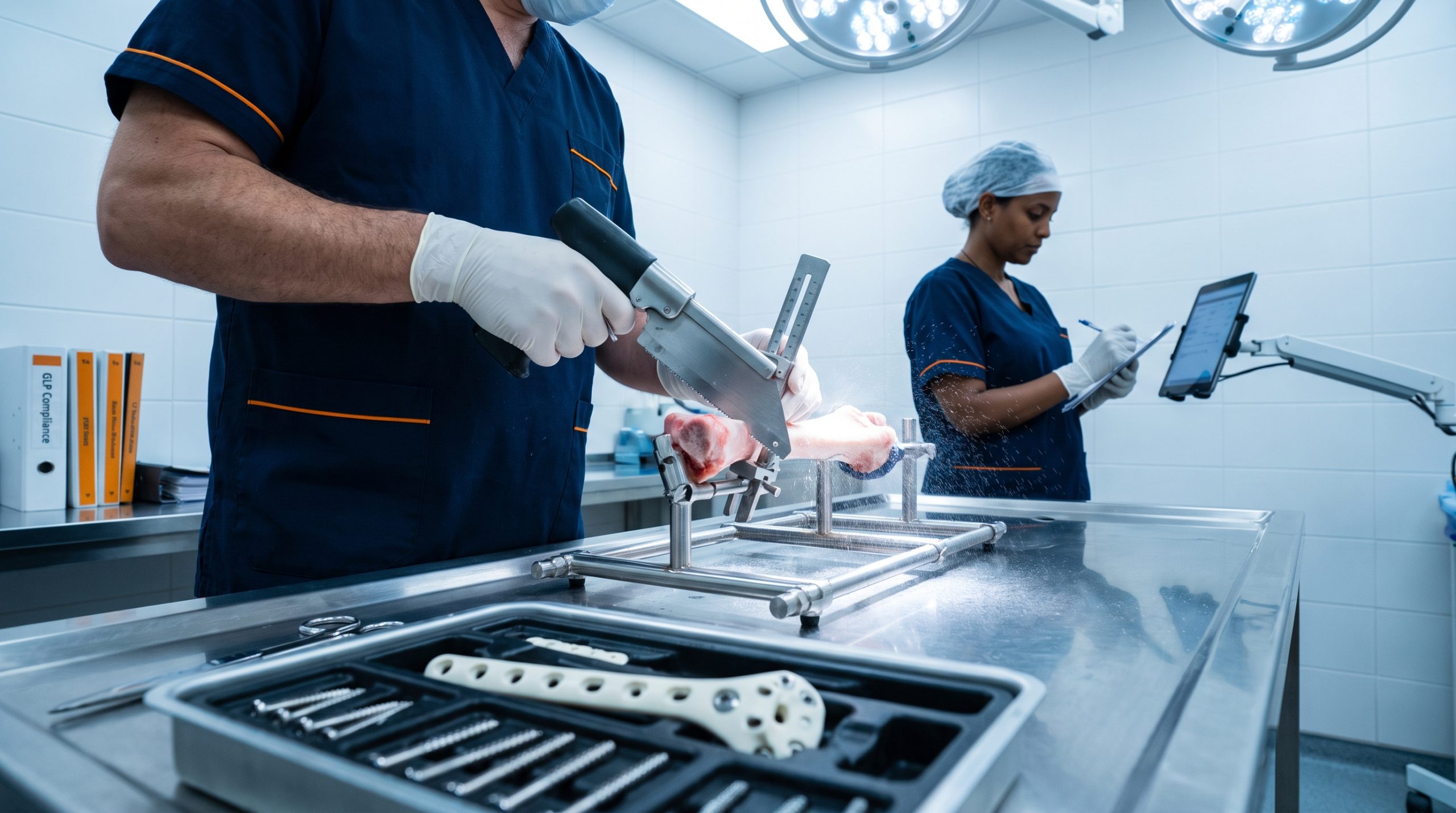
A critical-size defect is a bone void that will not bridge on its own within the observation period, so any healing observed can be attributed to the tested material or technique. In pigs, such defects are typically created with calibrated trephines or oscillating saws in cortical, cancellous or calvarial sites, with dimensions validated in pilot work before the pivotal study. Periosteum management, irrigation, thermal control during osteotomy and stabilization of the surrounding bone are all determinants of defect behavior. Because swine bone regeneration is vigorous, defect size must be verified for the chosen breed, age and location rather than borrowed from another species. Empty-defect controls, contralateral comparisons and standardized fill technique are what make a critical-size design defensible to reviewers assessing biomaterials, scaffolds or osteoinductive agents.
Anatomical sites commonly used for orthopedic implant testing in pigs
Site selection follows the device. The proximal and distal femur and the proximal tibia are frequent choices for cancellous screw fixation, augmentation materials and osseointegration coupons, offering generous trabecular volume and straightforward surgical exposure. Tibial and femoral diaphyses serve plate, nail and osteotomy studies where cortical bridging under load is the endpoint. The humerus supports fixation and anchor testing, while the calvaria and mandible are used for membrane, granule and non-load-bearing regeneration work. Lumbar and thoracic vertebrae accommodate interbody cages, pedicle screws and vertebral augmentation. In each case the rationale rests on three points: comparability of bone quality to the human target site, the loading environment the orthopedic implants will face clinically, and the ability of the porcine bone model to tolerate the procedure with good welfare outcomes.
Breed, age, and weight: matching the animal to the clinical question
Domestic breeds grow quickly and reach substantial mass, which suits short follow-up and human-scale device geometry but complicates long studies as the skeleton changes. Miniature and Göttingen-type pigs grow more slowly and are generally preferred for follow-up beyond a few months, where stable weight and closed growth plates protect data integrity. Skeletally immature animals are appropriate when the intended patient population is pediatric or when growth-plate interaction is the research question; mature animals better represent adult trauma and degenerative indications. Housing capacity, imaging table limits, anesthesia logistics and handling safety at the projected terminal weight should be checked before the protocol is fixed. Aligning breed, age and weight with the target population is what gives a porcine bone model genuine translational value for orthopedic implants.
Key outcome measures for swine orthopedic implant studies
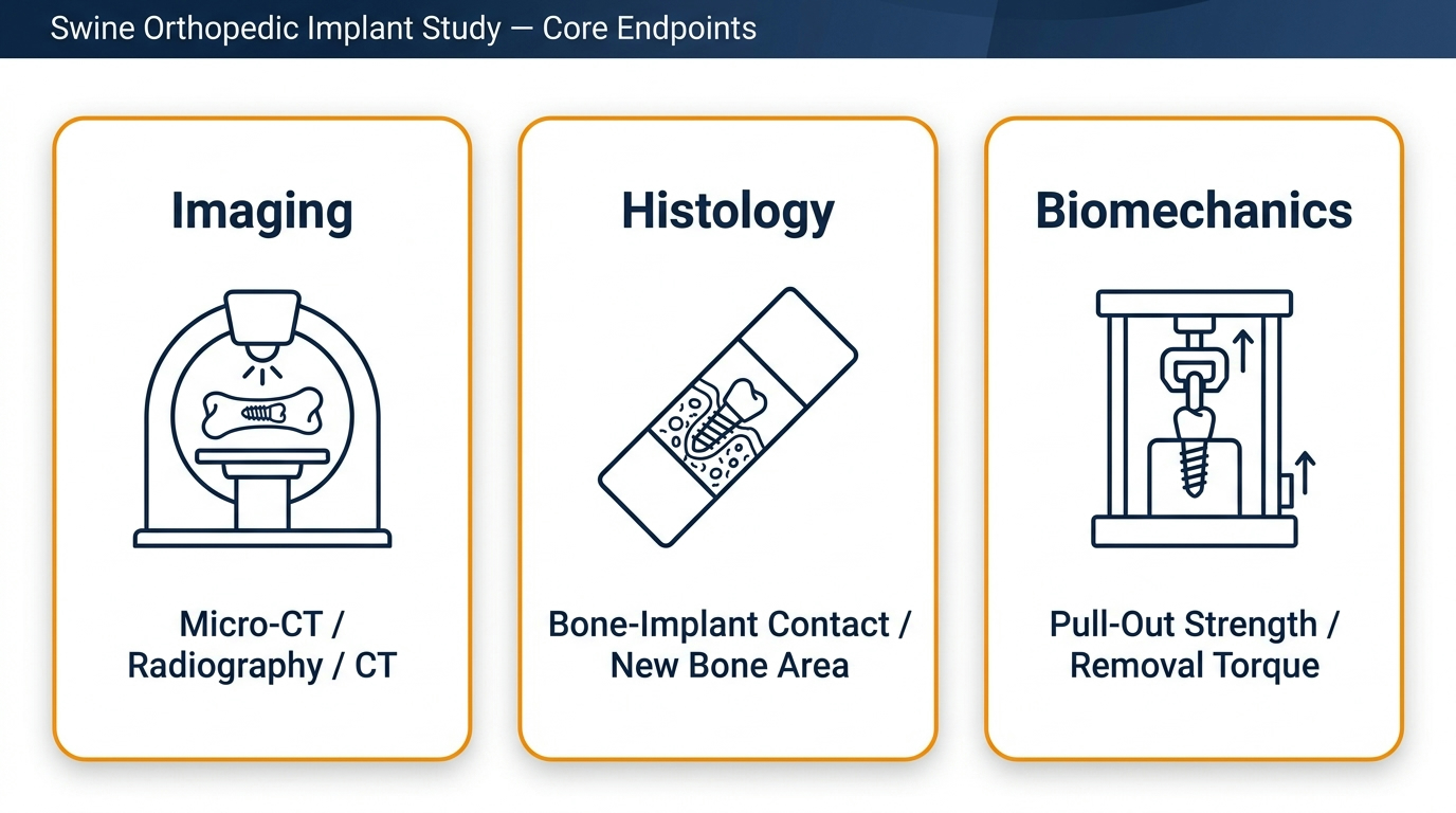
Robust swine orthopedic implant studies combine imaging, histology and mechanics rather than relying on any single modality. Published porcine work on titanium screws, for example, reports osseous integration assessed by micro-CT, mechanical testing and histology — a triad that has become the practical expectation for demonstrating osseointegration and swine bone regeneration. Endpoints should be pre-specified with defined regions of interest, thresholds and acceptance criteria.
Imaging techniques for orthopedic implant assessment
Radiography confirms implant position and gross alignment in the operating room and at interim timepoints. Fluoroscopy guides percutaneous placement in real time. Clinical CT provides three-dimensional callus and defect volume, while micro-CT of harvested specimens quantifies bone volume fraction, trabecular thickness, mineral density and peri-implant bone within the porcine bone model at resolutions relevant to bone-implant contact.
Histological and histomorphometric evaluation
Undecalcified resin embedding preserves the metal-bone interface; ground sections with toluidine blue or Stevenel/van Gieson staining allow quantification of bone-implant contact, new bone area, residual graft material, osteoid and marrow response. Decalcified paraffin sections support cellular and inflammatory scoring. Together they document maturation and biological tolerance underlying swine bone regeneration.
Biomechanical testing protocols
Pull-out and push-out tests quantify interfacial shear strength; removal torque characterizes screw integration; three- and four-point bending, axial compression and torsion assess healed segments. Consistent specimen alignment, potting method, crosshead speed and load-cell calibration are essential for comparable results across a porcine bone model cohort.
Methodology Box: Measuring Osseointegration for Regulatory Compliance
Osseointegration is defined as a direct structural and functional connection between ordered living bone and the surface of a load-bearing implant, and reviewers expect it to be demonstrated by converging evidence. In a pig model, that usually means micro-CT for peri-implant bone volume and architecture, histomorphometry for bone-implant contact percentage and new bone formation, and mechanical testing for interfacial strength or removal torque, all at pre-defined timepoints.
- Pre-specified sectioning planes and region-of-interest boundaries
- Blinding of assessors during histomorphometry and imaging analysis
- Pre-registered statistical comparisons across timepoints
- Calibration records and traceable specimen chain-of-custody
When these elements are planned before surgery rather than reconstructed afterwards, data supporting orthopedic implants tends to survive regulatory scrutiny with far fewer information requests.
What regulatory considerations apply to pig orthopedic implant testing?
Non-clinical safety studies intended to support a submission are normally conducted under Good Laboratory Practice, and the study plan should reflect regulatory expectations from the outset. The FDA guidance on general considerations for animal studies intended to evaluate medical devices addresses model justification, study design, monitoring and reporting content. Ethical oversight is mandatory: an institutional animal care and use committee or, in Israel, approval under the national animal experimentation framework and its council, must review and authorize the protocol before work begins. Biocompatibility and local-effect expectations are shaped by the ISO 10993 series, including the part covering local effects after implantation, and European submissions are further informed by MDCG guidance on preclinical evidence. Documented GLP, ethical approval and justified model selection are the backbone of credible pig model orthopedic device testing for orthopedic implants.
What does GLP mean in large animal device testing?
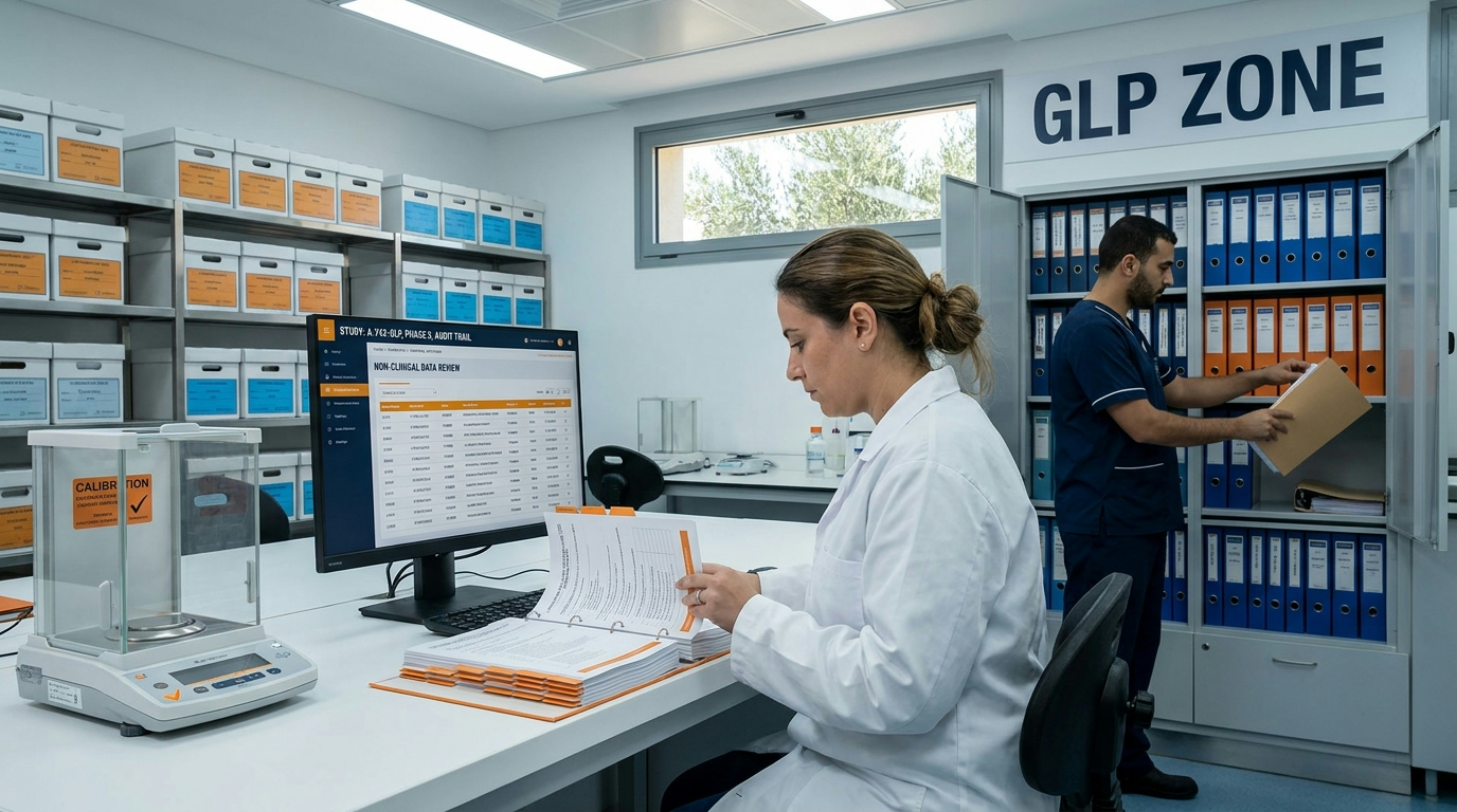
GLP is not a laboratory technique but a quality system governing how non-clinical safety studies are organized, performed, monitored, recorded, archived and reported, so that results are consistent, reproducible and traceable. In large animal device testing this touches almost everything: a named study director with defined authority, an approved and version-controlled protocol, standard operating procedures for anesthesia, surgery, imaging and necropsy, trained and documented personnel, calibrated equipment, characterized test articles, raw-data integrity rules and an independent quality assurance function. Animal identification, husbandry records, feed and water documentation, and deviation handling all become part of the evidence trail. Applied to pig model orthopedic device testing, GLP means that every claim in the final report can be traced back to a signed, dated, contemporaneous record — which is precisely what auditors and reviewers look for.
Good welfare is also good science. Pain and stress alter healing biology and confound bone outcomes — protocols that skip humane endpoints or standardized analgesia are not shortcuts, they are variance generators that ultimately weaken the very data the device team is trying to build.
— Adir Koreh, CEO, Biotech Farm Ltd.
How many animals are typically needed for a porcine orthopedic study?
Sample size for a porcine orthopedic study is driven by the primary endpoint, its expected variability, the effect size considered clinically meaningful, and the number of treatment and control groups multiplied by timepoints. Pilot or ex vivo data are invaluable for estimating variance, and paired designs — bilateral limbs or multiple implantation sites per animal — can substantially reduce animal numbers when independence assumptions are handled correctly. Attrition allowance for anesthetic risk, infection or fixation failure should be stated explicitly. The reduction principle within the 3Rs obliges sponsors to use the minimum number consistent with a statistically interpretable answer, and ethics committees will ask how that number was derived. Early involvement of a biostatistician usually protects both welfare and budget in pig model orthopedic device testing, because an underpowered study wastes animals as surely as an oversized one.
How long does fracture healing take in a pig model?
Timelines vary with defect type, fixation stiffness, anatomical site, and above all animal age. In young, rapidly growing pigs, early callus can be radiographically evident within two to three weeks, with cortical bridging in a stabilized osteotomy commonly assessed between four and twelve weeks. Cancellous osseointegration endpoints are often collected at four, eight and twelve weeks, while remodeling and material resorption questions may require six months or longer. A pig fracture healing model studying critical-size defects usually needs longer observation, since the point of the design is that healing does not occur unaided. Interim imaging at defined intervals, rather than a single terminal look, is what reveals the trajectory of swine bone regeneration and allows a sponsor to distinguish delayed healing from genuine non-union.
Complications to Anticipate in Swine Orthopedic Surgery Studies
Surgical site infection, implant loosening or migration, peri-implant fracture, hardware failure at screw-plate junctions, delayed union or non-union, seroma, and skin irritation over subcutaneous hardware are all documented complications. Mitigation begins pre-operatively: sterile technique, appropriate antibiotic and analgesia protocols, implant sizing verified on bench bone, and fixation stiffness matched to the animal’s expected activity. Predefined humane endpoints and a clear escalation pathway to the attending veterinarian protect both the animals and the integrity of the study. Transparent reporting of complications strengthens, rather than weakens, a preclinical dossier.
Mistakes that quietly undermine a porcine implant program
The most common error is retrofitting endpoints after surgery, which leaves no defensible statistical plan. A second is borrowing defect dimensions from another species without pilot verification, producing defects that heal spontaneously and erase the treatment signal. Third is ignoring growth: an animal that doubles in weight during follow-up changes the loading environment mid-study. Fourth is under-specifying histology — decalcified sections cannot answer bone-implant contact questions at a metal interface. Fifth is neglecting surgical rehearsal, so instrumentation problems consume the first animals. Finally, documentation gaps: uncalibrated equipment, unsigned records or unversioned protocols can invalidate otherwise excellent science. Each of these is preventable at the planning table, which is why an experienced study team and a written pre-study alignment meeting are worth more than any single piece of equipment.
A development scenario: from prototype screw to submission-ready evidence
Consider a company with a novel surface-treated cancellous screw. The output is a coherent narrative — bench, pilot, pivotal — that reviewers can follow. This staged logic is the practical shape of well-run pig model orthopedic device testing.
Phase 1: Ex Vivo Bench Testing
Insertion torque and pull-out strength measured in harvested porcine femora to confirm thread geometry and validate instrumentation before any live animal is enrolled.
Phase 2: Non-GLP Pilot Study
A small cohort refines surgical approach, implant site, imaging protocol and timepoints while confirming the model behaves as expected.
Phase 3: GLP Pivotal Study
Pre-specified micro-CT, histomorphometry and mechanical endpoints at multiple timepoints with a comparator control, locked analysis plan and signed final report.
What belongs in an animal study report for medical device submission?
A complete animal study report opens with objectives, the regulatory context and a justification of species and model selection. It then documents test and control articles with identification and traceability, animal details, husbandry, anesthesia and analgesia, the surgical procedure with operative findings, and the full schedule of observations. Results must present imaging, histopathology and biomechanical data with the pre-specified statistical analysis, individual animal data appendices, and honest disclosure of deviations, complications, unscheduled deaths and negative findings. Discussion should interpret translational relevance and limitations, followed by conclusions tied strictly to the data. Signatures of the study director, pathologist and quality assurance unit, plus archiving statements, complete the package. Reports written to this standard support a medical device submission efficiently because they demonstrate GLP discipline on every page.
Can human-sized orthopedic implants be tested effectively in pigs?
Yes — and this is one of the main reasons sponsors move from rodents to swine. Porcine femora, tibiae and vertebrae accommodate clinically dimensioned plates, nails, screws, anchors and interbody devices without scaling down the prototype, so surgeons evaluate the actual instrument set, the actual insertion sequence and the actual fit. Body mass in the 60–120 kg range delivers loading conditions relevant to adult patients, allowing assessment of primary stability, subsidence, screw purchase and fatigue-related loosening. Programs built around Swine Models Biomedical Research therefore frequently center on human-sized orthopedic implants, because testing the production device rather than a miniature analogue removes an entire layer of translational uncertainty from pig model orthopedic device testing and shortens the path to clinical evaluation.
Typical deliverables from a porcine implant study
Sponsors should expect a defined document and data package rather than a verbal summary. Standard deliverables include the approved study protocol with amendments, ethics approval documentation, surgical and anesthesia records, interim reports, raw and processed imaging sets (radiographs, fluoroscopy captures, CT and micro-CT datasets), histology slides with digital scans and a pathology report, biomechanical test curves and derived parameters, the statistical analysis, and a signed final report. For comprehensive PIG – Large animal models for R&D projects, deliverables often extend to procedural video for training and design-history purposes, explanted device analysis, custom endpoint development and confidentiality arrangements protecting intellectual property. Clarifying deliverables in the quotation stage prevents disputes later and ensures the porcine implant study output aligns with what the regulatory file actually needs for orthopedic implants.
Mapping sponsor needs to practical support at a large animal facility
Different sponsors arrive at different stages, and useful support looks different in each case. The table below maps common business needs to what a well-equipped facility can contribute in practice, without implying that any single arrangement fits every program.
| Sponsor need | How a full-service facility helps in practice |
|---|---|
| Concept still evolving | Scientific escort and brainstorming sessions in an interactive conference setting before the protocol is locked |
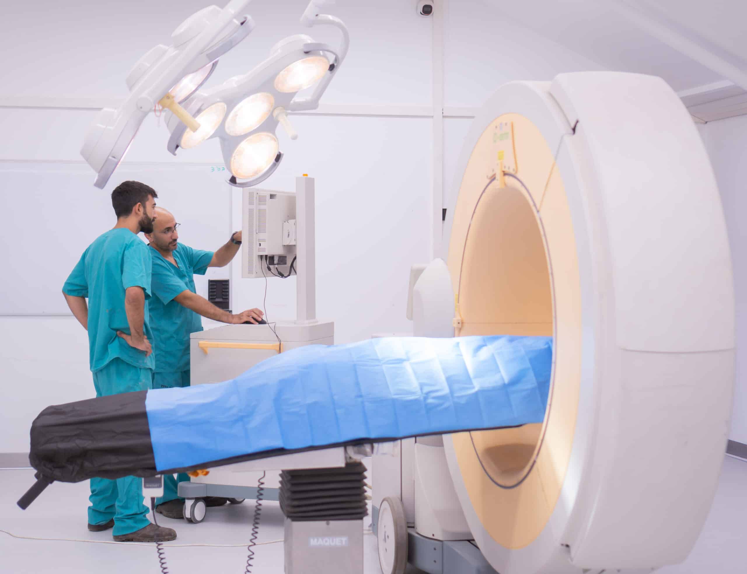
| Imaging-heavy endpoints | On-site C-Arm fluoroscopy, high-definition and cardiac ultrasound, OCT and surgical microscopy in the same surgical suite |
| Minimally invasive technique | 4K laparoscopic towers and senior surgeons experienced in percutaneous and arthroscopic approaches |
| Regulatory documentation | Protocol drafting aligned with GLP-style documentation, ethics submission support and structured final reporting |
| Animal welfare assurance | Spacious animal housing, trained handling, analgesia protocols and 3Rs-driven design (replacement, reduction, refinement) |
| Israel-based development teams | Local access for repeated site visits, hands-on surgical rehearsal and same-day sponsor observation |
Partnering with Biotech Farm for Your Pig Model Orthopedic Device Testing
Working with a dedicated large animal facility changes the tempo of a program. At Biotech Farm the team brings more than three decades of experience in leading and managing research, combined with Preclinical Research And Development Services spanning orthopedics, cardiology, ophthalmology, wound healing and metabolic platforms. Two large, fully equipped surgery rooms, advanced imaging, a professional veterinary and surgical crew, and a comfortable animal house support studies from feasibility through pivotal reporting. Transparency in collaboration and well-documented procedures mean sponsors see the data as it is generated rather than only at the end.
Frequently asked questions about porcine orthopedic studies
Ready to define the right porcine model for your device?
Which endpoint will your reviewers scrutinize first — osseointegration, biomechanical stability, or the completeness of your documentation? If you are scoping a study and want to pressure-test the design before committing animals and budget, our surgical, veterinary and scientific team is available to review your objectives, propose a fitting porcine bone model, and outline realistic timelines and deliverables. Bring your device drawings, intended indication and target submission pathway so the discussion can be concrete from the first meeting.