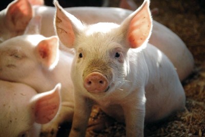
With more than 20 years of hands-on practice in large animal model setup, our team at BIOTECH FARM Ltd. has supported urological device developers and surgical teams through every stage of preclinical validation. When a ureteroscope, catheter, or stent is about to meet human tissue for the first time, the pig model is where it proves itself — and a poorly designed study wastes that opportunity. This guide explains how the model works, when to use ex vivo versus in vivo setups, and how to design a study that produces answers, not just activity.
Expert Insight: The most common reason a pig model study underdelivers is not the model — it is skipping the characterization phase. Teams that define endpoints, model type, and success criteria before the first organ is prepared consistently get decision-grade data. Teams that arrive with only a device in hand get videos and anecdotes.
What Is a Pig Model for Urological Procedures and Who Is It Suitable For?
A pig model for urological procedures is a research and training platform that uses porcine anatomy, either in vivo (in a living animal) or ex vivo (isolated organs), to study, validate, and rehearse interventions on the urinary tract. Pigs are chosen because the size, structure, and tissue behavior of their kidneys, ureters, bladder, and urethra are close enough to the human equivalent to support realistic work with clinical instruments. Teams that develop endoscopes, catheters, stents, and access devices, as well as surgeons who need to refine technique before clinical exposure, both benefit from this model.
The scope of a pig model can be narrowed to a single organ, such as the kidney for percutaneous access work, or expanded to the full urinary tract for transurethral procedures that pass from the urethra through the bladder and into the ureter. The choice depends entirely on the research question, the endpoints that matter, and the stage of development of the device or technique being evaluated.
How Do In Vivo, Ex Vivo, and Bench Models Differ?
An in vivo model involves a living, anesthetized animal. It preserves full physiology: blood flow, bleeding response, tissue perfusion, healing, and systemic reactions. This makes it the right setting when safety, biocompatibility, or long-term tissue response is part of the question. An in vivo model also allows imaging during the procedure, such as fluoroscopy and ultrasound, in conditions that closely mimic an operating room.
An ex vivo model uses organs harvested after sacrifice, typically kidney with ureter attached, or bladder with urethra, and keeps them functional in a controlled setup with perfusion or fluid filling. The researcher controls pressure, flow, temperature, and filling rate with engineering precision, and can repeat the same maneuver many times under identical conditions. A bench model, by contrast, is non-biological: simulators, silicone phantoms, and artificial tubing. It is useful for very early device concept checks and basic coordination training, but it cannot reproduce tissue compliance, friction, or mucosal interaction, which is exactly where porcine tissue adds value.
Why Pigs Rather Than Smaller Animals?
Rodents and rabbits have long been used in pharmacology, but their urinary tract is simply too small for clinical-diameter instruments. A pig ureter accepts a standard ureteroscope, a pig bladder holds realistic volumes, and a pig kidney has a collecting system that can be entered, navigated, and stented with the same tools a surgeon would use on a patient. This single factor changes the nature of the data: instead of scaled-down approximations, the researcher measures real device performance in real tissue dimensions.
Beyond instrument compatibility, porcine tissue properties matter. The mucosa, muscle layers, and connective tissue of the urinary tract behave mechanically like human tissue during manipulation, dilation, and perforation. That is why the porcine model is widely described as one of the most accurate representations of the human kidney and urinary tract for surgical simulation and research, as reviewed in a recent MDPI review of the porcine model in urological research and training.
How Similar Is the Pig Urinary Tract to the Human One?
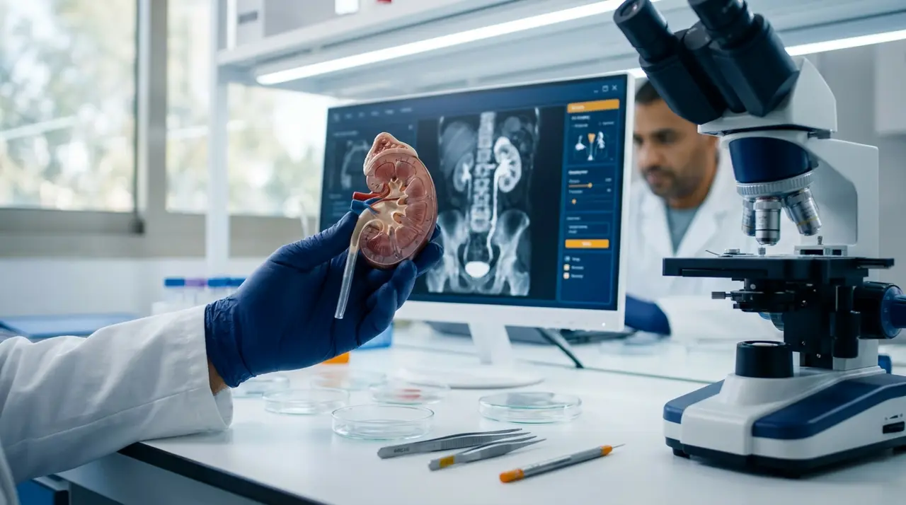
The similarity is high enough to support a very wide range of preclinical and training protocols, but it is not absolute, and honest protocol design accounts for the differences. In the upper tract, the number and orientation of renal calyces, the length and diameter of the ureter, and the anatomy of the ureterovesical junction can all differ from the human pattern. These differences affect how easily an endoscope passes, the angles available for manipulation, and the kinematics of stone work or biopsy within the collecting system.
In the lower tract, bladder capacity, trigone anatomy, and especially the male urethral curvature can make catheterization and cystoscopy more demanding than in a human patient. Image-guided studies of swine anatomy have documented these procedural limitations explicitly, which is valuable: knowing where the model diverges tells you which findings transfer to the clinic and which are artifacts of the anatomy. A well-designed protocol therefore defines what is measured, not only what is performed, so that anatomical confounders are separated from device or technique performance.
Which Procedures Can Actually Be Performed on the Pig Urinary Tract?
The practical catalogue is broad. On the upper tract, teams routinely perform ureteroscopy (flexible and semirigid), retrograde intrarenal surgery, percutaneous renal access, nephrostomy placement under ultrasound or fluoroscopy guidance, and percutaneous nephrolithotomy access training. Guidewire handling, tract dilation, sheath placement, and stent deployment are all rehearsed and measured. On the lower tract, cystoscopy, catheterization, bladder filling and emptying protocols, urodynamic measurement, and mucosal interaction studies are all feasible.
Functional endpoints can be attached to any of these: intraluminal pressures, irrigation and drainage flow rates, time to target, number of attempts, success rates, and visible or histological tissue trauma. Even complex surgical scenarios such as kidney transplantation have been developed and optimized in pigs, including a reproducible low-weight miniature pig model described in this transplantation study, and a step-by-step heterotopic kidney allotransplantation model detailed by the International Journal of Organ Transplantation Medicine.
What Is a Porcine Urinary Tract Model and What Is Tested In It?
A porcine urinary tract model treats the whole system, kidneys, ureters, bladder, and urethra, as a connected platform. This is important when the procedure itself crosses anatomical boundaries: a transurethral stent placement passes the urethra, enters the bladder, negotiates the ureterovesical junction, and terminates in the renal pelvis. A full-tract model allows each of these stages to be simulated and timed separately, or the whole chain to be evaluated end to end.
What is actually tested falls into three families: navigation performance (can the device reach the target, and how quickly), therapeutic action (does the treatment step work, whether that is stone fragmentation, dilation, or drug delivery), and completion quality (is the stent correctly positioned, is drainage established, is the mucosa intact). Facilities such as Biotechfarm’s swine biomedical models program support exactly this kind of staged protocol design, where the same session can yield both training metrics and device performance data.
When Is a Pig Bladder Model the Right Choice?
A bladder-focused model is preferred whenever the research question centers on pressure, volume, drainage, or the interaction between a device and the bladder wall. Typical applications include urinary catheter performance testing, where drainage rate, patency of the eyelets, and blockage behavior are measured; urodynamic studies, where pressure-volume curves during filling and emptying are recorded; and mucosal trauma assessment, where repeated catheterization is evaluated through cystoscopy and histology.
The ex vivo version of this model offers exceptional experimental control. A validated isolated pig bladder can sustain filling and emptying protocols for pressure-volume measurement, and perfused versions have been used to study ischemic decompensation and even neurophysiological signals from afferent nerves, with urodynamic catheters integrated into the setup. This means a bladder model can go far beyond basic flow metrics and deliver physiological endpoints in a fully standardized environment.
Why the Pig Kidney Model Is Central to Endourological Research
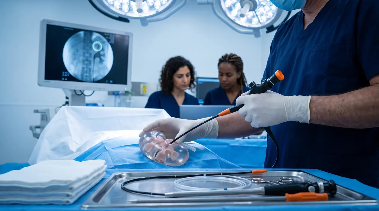
Kidney-focused work dominates endourology for an obvious reason: most innovation targets the collecting system. Accessing the kidney percutaneously, navigating the calyces, and deploying instruments within a confined, curved anatomy are the hardest skills and the riskiest device requirements. The porcine kidney allows all of this to be rehearsed with real tissue feedback: resistance during puncture, guidewire behavior across the infundibula, and the feel of sheath advancement.
Scenario design is what turns the model into a measurement tool. A target can be defined within the collecting system, for example a calyx or a simulated stone, and performance is then quantified as time to target, number of puncture attempts, fluoroscopy time, and complication events. Biological models based on porcine kidneys have been shown to be effective for renal access training and have been integrated into structured training programs with measurable skill improvement.
Urinary Tract Model Versus Lower Urinary Tract Model: A Practical Comparison
The distinction sounds subtle but it changes the experiment. A full urinary tract model includes the upper system and enables transurethral passage all the way to the renal pelvis, which is essential for stenting and ureteral access studies. A lower urinary tract (LUT) model isolates the bladder and urethra, which allows an even more controlled setup: simulated abdominal pressure, defined filling rates, and measured emptying volumes, all while the device sits in the exact anatomy it will occupy clinically.
An ex vivo porcine LUT model has been developed specifically to evaluate urinary catheter performance, including direct observation of how the catheter eyelets interact with the bladder mucosa, alongside hydrodynamic parameters. For a catheter developer, that combination of visual and quantitative data in one session is hard to obtain in any other way.
Choosing Between Ex Vivo and In Vivo: A Decision Matrix
The choice should be driven by endpoints, not by habit or cost alone. The table below maps common use cases to the appropriate model type:
| Use Case | Recommended Model | Rationale |
|---|---|---|
| Early device concept validation / proof of concept | Ex vivo | Fast iterations, low cost per repetition, controlled conditions |
| Surgical technique refinement and repetitive training | Ex vivo | Many repetitions on identical anatomy, no survival burden |
| Physiological response and systemic effects | In vivo | Requires perfusion, bleeding response, and whole-animal physiology |
| Safety and biocompatibility evaluation | In vivo | Healing, inflammation, and systemic reaction can only be assessed in a living animal |
| High-fidelity simulation of complex full procedures | In vivo | Full anatomical continuity and imaging conditions, though ex vivo suits specific task drills |
| Advanced urodynamic measurements | Ex vivo perfused | Instrumented setup with urodynamic catheters and precise flow control |
Flexible ureteroscopy training on isolated porcine kidneys has been validated as a realistic in vitro alternative, while in vivo porcine models support retrograde intrarenal surgery practice with the caveat that anatomical differences between pig and human must be factored into interpretation.
What Endpoints and Success Metrics Are Common?
For device testing, the standard metrics are placement accuracy, insertion and withdrawal forces, friction along the tract, blockage or occlusion events, device failures, and repeatability across uses. For procedure testing, the metrics shift to number of attempts, time to target, radiation or fluoroscopy time, and adverse events such as perforation, bleeding, or extravasation. Both families of metrics benefit from standardized documentation and, ideally, video capture of each run.
Tissue-level endpoints add depth. Cystoscopy performed after the procedure, followed by pathology and histology, reveals mucosal trauma that is invisible during the operation itself, as demonstrated in an in vivo pig study of intermittent catheterization. For stent development, the porcine model is considered highly suitable precisely because anatomical similarity lets encrustation, migration, and drainage behavior be observed in realistic conditions, and combined in vitro and in vivo methodologies are standard practice in this field.
Where Device Testing and Surgical Training Part Ways
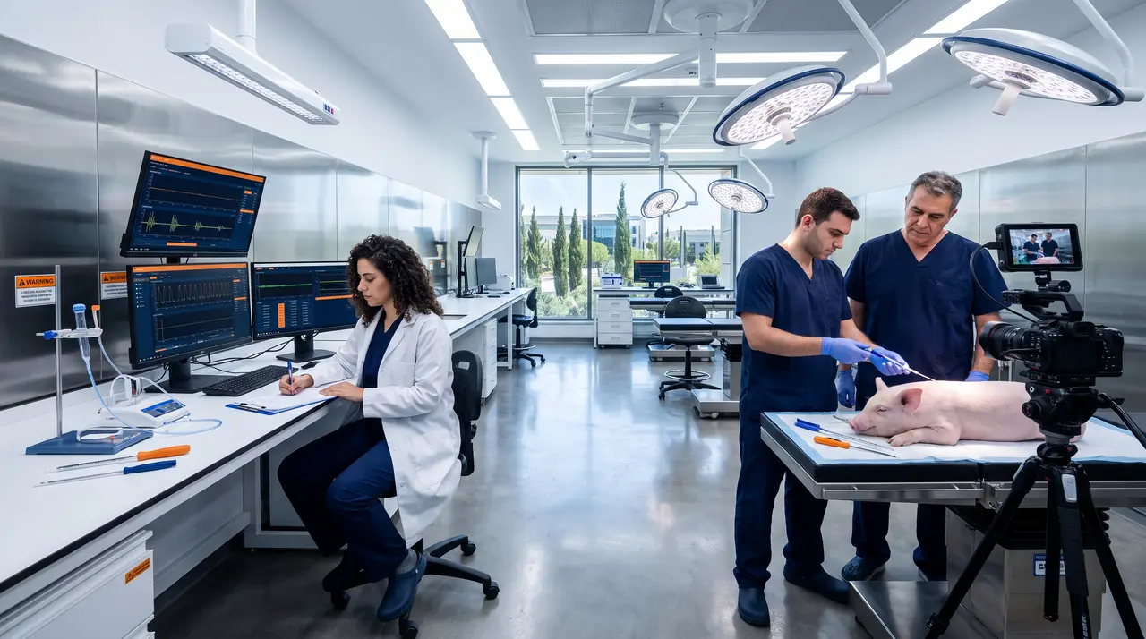
Although the same facility and the same animals can serve both purposes, the protocol logic differs. Device testing is a product development activity: it demands rigorous experimental design, pre-defined quantitative endpoints, standardization across repetitions, and documentation quality that can eventually support regulatory submission. The question is whether the device performs as intended and how it fails when it fails.
Surgical training is a human factors activity. The unit of analysis is the trainee, not the device, so the protocol needs progressive difficulty levels, scenario variety, and structured feedback, often combining video review with objective metrics such as time, attempts, and fluoroscopy dose. Ex vivo porcine models for ureteral stent placement training, for example, track exactly these operational metrics and demonstrate gains in confidence and knowledge. Validated endourology curricula frequently combine several model types, using porcine tissue for the stages where realism matters most, such as ureteroscopy and management of urological injuries.
Skipping the characterization phase is the most common cause of a study that produces activity but not answers.
— BIOTECH FARM Ltd. Research Team
How Does a Commercial Pig Model Project Actually Run?
A well-run commercial engagement begins with characterization: what is the product or procedure, who is the intended user, and what would count as success. This phase determines whether the work is best done ex vivo or in vivo, on kidney, bladder, or the full tract, and which measurements will answer the sponsor’s question. Skipping this step is the most common cause of a study that produces activity but not answers.
The execution phase covers animal selection, protocol finalization, ethics approval if required, scheduling of imaging and surgical resources, and standardized data capture across every run. The delivery phase then packages raw data, analysis, and conclusions into a report the sponsor can act on, whether that decision is design iteration, a move to in vivo work, or preparation for regulatory conversations. Providers that integrate surgery rooms with fluoroscopy, high-definition ultrasound, and laparoscopic towers under one roof reduce handoffs and keep the data chain consistent.
Our Methodology at a Glance
- Characterization: defining the product, intended user, success criteria, and model type before any preparation begins
- Execution: animal selection, ethics compliance, imaging resources, and standardized data capture on every run
- Delivery: raw data, analysis, and actionable conclusions packaged to support design iteration or regulatory conversations
What Regulatory and Ethical Rules Apply in Israel?
Research involving animals in Israel operates under the Animal Welfare Law (Experiments on Animals), 1994, which defines the legal framework for all animal experimentation. The Ministry of Agriculture and Rural Development’s ethics committee for animal experimentation applies principles that include demonstrating that no alternative exists, using the minimal number of animals necessary, and institutional and veterinary responsibility for welfare throughout the study. Any project at a facility in Israel must be planned around these approval requirements from the outset.
Reporting quality is governed by guidelines such as ARRIVE 2.0, which cover rationale for the chosen model, experimental design, transparency, and reproducibility. Sponsors who intend to use preclinical data in later regulatory discussions benefit when the provider already works to these standards, because the documentation burden is handled during the study rather than reconstructed afterward. When a project also involves pharmacokinetic or drug delivery components alongside the urological work, facilities with combined capabilities, such as Biotechfarm’s drug delivery models and PK determination services, allow both data streams to be collected under one protocol and one ethical framework.
Frequently Asked Questions
Which of these questions matters most for your current program — a device approaching validation, a technique awaiting refinement, or a training need with measurable goals? If you are weighing whether an ex vivo kidney setup, a full urinary tract model, or an in vivo protocol fits your endpoints, the team at Biotechfarm can help map the options and design a study that answers your specific question.