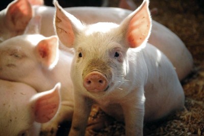
Preclinical cardiovascular work succeeds or fails long before the first animal is anesthetized — it is decided in the study design. With more than 20 years of hands-on large animal model experience, Adir Koreh and the Biotech Farm team have run porcine cardiovascular programs across coronary, structural heart, electrophysiology, and heart failure platforms. Pig model cardiovascular device testing sits at the exact point where an engineering concept meets living hemodynamics: pressures, pulsatile flow, coagulation, arrhythmia thresholds, and healing biology. This guide walks through how a defensible, regulator-ready program is assembled, which metrics matter, and where studies commonly lose time.
Exclusive Insight: Why Sequencing Matters More Than Species
The most efficient programs never treat in vivo work as the first step. Ex vivo runs narrow the design space and burn no animal lives; in vivo studies then confirm the surviving configuration. This sequencing is the most defensible expression of scientific economy — it reduces the number of animals required to reach a decision while still producing regulator-ready data.
Why Pigs? The Anatomical and Physiological Advantage in Cardiac Research
The porcine heart is remarkably close to the human heart in mass-to-body ratio, chamber geometry, valve architecture, great vessel dimensions, and conduction system behavior. Coronary distribution is largely left-dominant, with vessel diameters in the range routinely treated in interventional cardiology, which is why pig coronary artery models accept clinical guide catheters, guidewires, balloons, and stents with only modest procedural adaptation. Comparative reviews describe the domestic pig’s coronary system as functioning in a fashion similar to human coronary arteries, supporting its use as a translational cardiovascular platform (representative porcine model for human cardiovascular research). Practical procedural guides also detail the anesthetic, antiarrhythmic, and anticoagulation adjustments required for angiography and PCI in swine (porcine coronary angiography and PCI guide). Those adjustments are exactly where hands-on experience separates a smooth study day from a lost animal.
What Exactly Does a Pig Model Cardiovascular Device Testing Program Include?
Defined simply, pig model cardiovascular device testing is a structured set of preclinical experiments in swine designed to evaluate safety, deliverability, deployment accuracy, acute performance, tissue response, and overall clinical feasibility of a cardiovascular device under human-like conditions. Regulatory guidance for device animal studies emphasizes structured design, defined objectives, monitoring, evaluation criteria, and complete reporting rather than descriptive observation (FDA general considerations for animal studies). In practice, deliverables include an approved protocol and study plan, procedural records, angiographic and intravascular imaging files, hemodynamic datasets, necropsy findings, histopathology with quantitative morphometry, and an integrated final report. At Biotech Farm, that package is built with the sponsor’s regulatory strategy in mind, so pig heart device testing data arrives in a form that engineering, quality, and clinical teams can all use.
In Vivo or Ex Vivo: Which Porcine Setup Answers Your Question?
An in vivo pig cardiovascular model is the right choice when the question involves living biology: device deployment in a beating heart, thrombogenicity under real coagulation, arrhythmia induction, vessel wall injury, neointimal healing, systemic hemodynamic response, or chronic durability. An ex vivo pig cardiovascular model — isolated hearts, explanted valves, or perfused vessel segments in a controlled circuit — excels where repeatability and parametric control matter more than biology: flow visualization, pressure gradient mapping, crimping and expansion mechanics, anchoring force, and rapid design iteration. Many efficient programs use both.
Inside the Swine Cardiac Model: Heart-Specific Device Evaluation
Where the coronary model looks at vessels, the swine cardiac model looks at the heart as a pump and an electrical organ. Studies here address ventricular function, valve competence, septal anatomy, conduction and rhythm stability, chamber pressures, and the response of a compromised myocardium to therapy. Transcatheter platforms rely heavily on this model because access route, sheath compatibility, annular sizing, anchoring behavior, and paravalvular sealing can only be judged inside a moving, pressurized heart. Echocardiography, fluoroscopy, and invasive pressure recording run in parallel during the procedure, so deployment events are correlated in real time with functional consequences. Reviews of swine as a platform for cardiac surgery and device development describe this combination of procedural realism and measurable physiology as the core reason pigs remain a default choice (pigs as models to test cardiovascular devices).
A Scenario: A Coronary Stent Program from Feasibility to Chronic Follow-Up
Week One: Acute Feasibility
Femoral or carotid access, engagement of the LAD and circumflex, deliverability through tortuosity, and acute recoil measured by quantitative coronary angiography in two to three animals.
Pivotal Chronic Arm
Multi-vessel implantation with defined stent-to-artery overstretch ratios, imaging follow-up at 28 days for early healing and 90 days for late lumen loss and inflammation resolution.
Terminal Pathology
Explanted vessels undergo resin embedding, sectioning, and morphometry — injury score, inflammation score, neointimal area, percent area stenosis, and endothelialization grade.
This is what a mature pig coronary artery model workflow looks like in practice — every stage produces the evidence base that regulators expect.
Normolipemic Against Atherosclerotic Substrates: Matching the Disease State
A healthy juvenile pig heals fast and rarely develops human-like plaque, which is ideal for biocompatibility questions and misleading for plaque-modification questions. Choosing the substrate is therefore a scientific decision with direct budget and timeline consequences. Diet-induced or familial hypercholesterolemia models take months to mature and cost more, but they are the only way to interrogate devices whose mechanism depends on lipid-rich or calcified tissue.
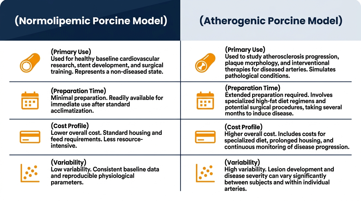
| Aspect | Normolipemic (healthy) model | Atherogenic / diseased model |
|---|---|---|
| Primary use | Healing response, neointima, biocompatibility, deliverability | Plaque interaction, calcium, drug uptake in diseased wall |
| Preparation time | Days to weeks | Several months of diet or induction |
| Cost profile | Lower, highly predictable | Higher, with husbandry and monitoring overhead |
| Variability | Low between animals | Moderate to high; requires larger groups |
| Typical endpoints | Injury/inflammation scores, endothelialization | Plaque burden, composition, lesion-level response |
Which Devices Are Actually Tested in a Porcine Cardiovascular Model?
The device range is broad. Intracoronary technologies include bare-metal and drug-eluting stents, bioresorbable scaffolds, drug-coated balloons, atherectomy and thrombectomy systems, and embolic protection. Structural heart work covers transcatheter aortic, mitral, and tricuspid valves, surgical prostheses, annuloplasty and repair devices, septal and appendage occluders, and shunt devices. Intracardiac and electrophysiology work includes leadless pacemakers, ablation catheters, mapping systems, and implantable sensors.
- Coronary stents, bioresorbable scaffolds, and drug-coated balloons
- Transcatheter aortic, mitral, and tricuspid valves
- Septal and appendage occluders, shunt devices
- Leadless pacemakers, ablation catheters, and mapping systems
- Vascular access hardware, closure devices, and grafts
Each class demands a different anatomical target, imaging plan, and follow-up horizon, which is why model selection precedes protocol writing rather than following it. For a broader look at how species and platforms are matched to vascular implants, see our discussion of stent testing animal models.
Model Selection Map: Matching Device Class to Porcine Platform
Selecting the right swine cardiac model is a matter of aligning the device’s mechanism of action with the anatomy and physiology that can actually stress it. Structural heart programs deserve particular attention, because growth rate in juvenile swine can alter annular dimensions across a long follow-up — a consideration explored further in our guide to the heart valve replacement animal model.
| Device class | Recommended platform | Core questions | Typical follow-up |
|---|---|---|---|
| Coronary stents/scaffolds | Coronary model, normolipemic or atherogenic | Deliverability, injury, neointima, endothelialization | Acute, 28, 90, 180 days |
| Transcatheter valves | Structural heart model, size-matched | Access, anchoring, gradients, paravalvular leak | Acute to 150+ days |
| Occluders and shunts | Septal / transseptal model | Positioning, stability, endothelial coverage | 30–180 days |
| Ablation/pacing systems | Electrophysiology model | Lesion depth, capture thresholds, rhythm safety | Acute to 90 days |
| Support/assist devices | Ischemic or heart failure model | Unloading, output, myocardial recovery | Model-dependent |
Heart Failure and Ischemia Models: When the Substrate Must Be Sick
Devices intended for failing hearts cannot be judged in healthy ones. Porcine ischemia and heart failure models are induced through several validated routes: transient or permanent balloon occlusion of a coronary branch to create infarction, ameroid constrictors or specialized flow-limiting stents to produce chronic ischemia with collateral development, microembolization, or rapid pacing to generate dilated cardiomyopathy. Each route yields a different phenotype — regional wall motion abnormality, global dysfunction, or ischemic burden with preserved viability — and the choice should follow the device’s claimed mechanism.
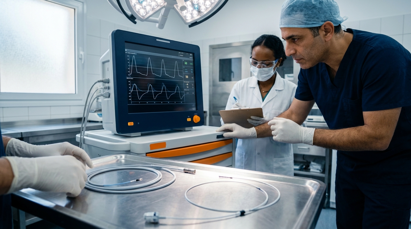
Endpoints typically combine ejection fraction and strain by echocardiography, invasive pressure-volume analysis, infarct size and perfusion by imaging, viability testing, and terminal histology. Because these models carry higher procedural risk and mortality, group sizing and interim monitoring plans must be defined before the first animal enters the protocol.
Endpoints and Measurements That Regulators Actually Read
A study is only as strong as its measurable outcomes. Well-constructed pig model cardiovascular device testing defines primary and secondary endpoints in the protocol, specifies the analysis method for each, and pre-registers the time points. The three pillars are imaging, hemodynamics, and pathology, and they should be planned as one integrated dataset rather than three separate reports.
Methodology Overview: The Integrated Dataset
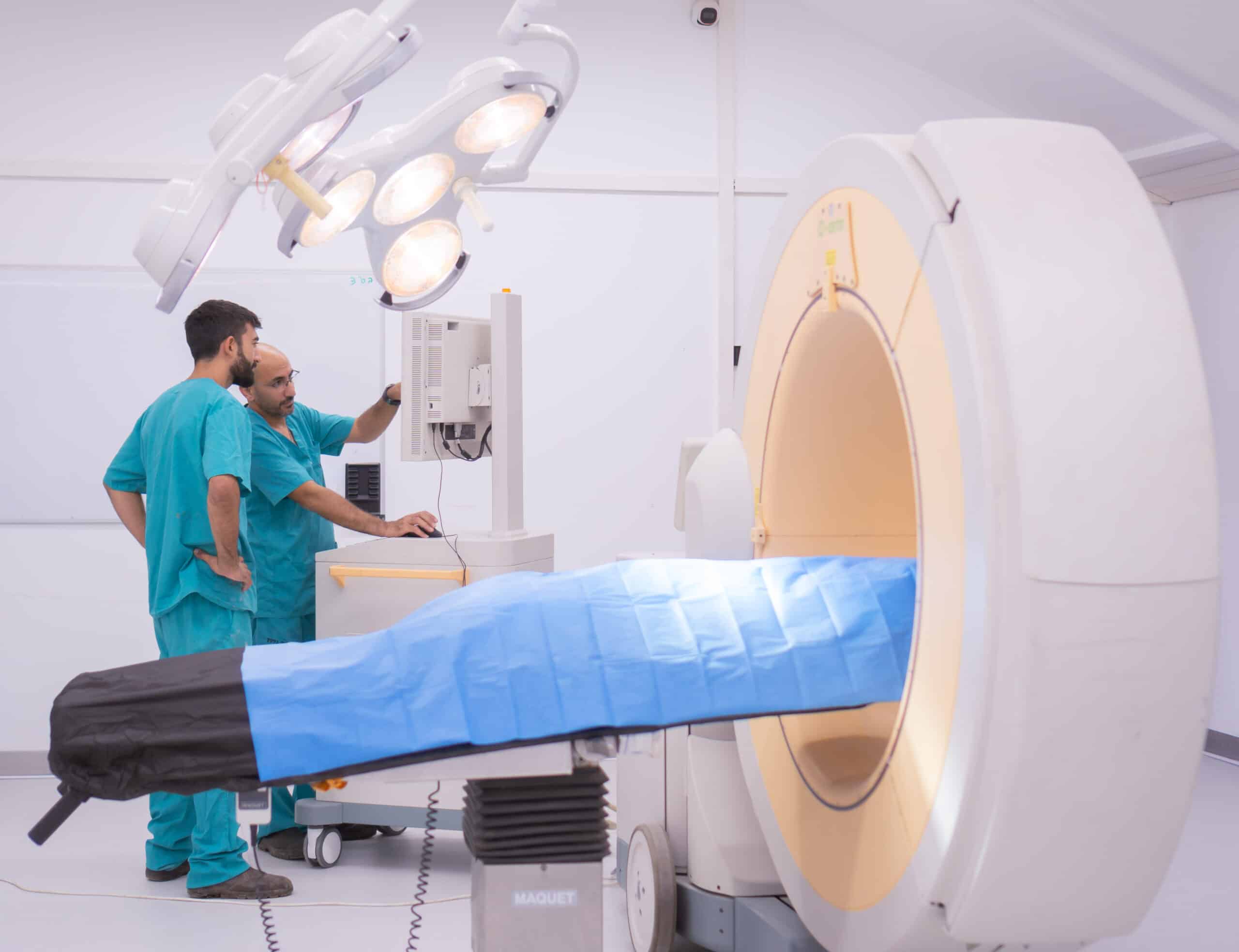
Imaging — Quantitative coronary angiography for lumen diameter, acute gain, and late loss; IVUS for apposition and remodeling; OCT for strut coverage; echocardiography for chamber and valve function; MRI for perfusion and infarct characterization. Biotech Farm’s surgical suites house a C-Arm, high-definition and cardiac ultrasound, OCT, and 4K laparoscopic towers in one room during one anesthesia session.
Hemodynamics — Aortic and left ventricular pressures, end-diastolic pressure, dP/dt, transvalvular gradients, cardiac output, and pressure-volume loop analysis with conductance catheters for load-independent indices such as end-systolic elastance.
Pathology — Macroscopic necropsy, high-resolution radiography of implants, resin or paraffin processing, and blinded scoring for injury, inflammation, fibrin deposition, and endothelialization, correlated back against the earlier imaging time points.
Consistency matters more than sophistication: identical anesthetic depth, ventilation settings, fluid status, and catheter positioning across animals will do more for data quality than any single advanced index acquired under variable conditions.
— Adir Koreh, CEO, Biotech Farm Ltd.
Common Mistakes That Derail Porcine Cardiovascular Studies
The recurring failures are rarely exotic. Animals are selected by age rather than by weight and target vessel diameter, producing oversized or undersized deployment. Antiplatelet and anticoagulation regimens are started too late, creating acute thrombosis that is a protocol artifact rather than a device property. Ventricular fibrillation during coronary manipulation is anticipated too casually, without amiodarone or lidocaine prophylaxis and immediate defibrillation readiness.
Warning: Growth Distorts Sizing
Growth of juvenile swine over a 90- or 180-day follow-up is frequently ignored, distorting valve sizing and vessel-to-device ratios. Imaging views that change between time points make quantitative comparison impossible. Each of these is preventable with a pilot phase, written procedural checklists, and a study director who has personally run the procedure many times before.
Variability and Reproducibility: How Many Animals, and Why
Biological variability is not noise to be apologized for; it is a parameter to be managed. Reproducibility improves when animals come from a defined source with narrow weight ranges, when anesthesia and analgesia protocols are standardized, and when device assignment is randomized across vessels and animals to neutralize position effects. Group size should follow a power calculation based on the expected effect size of the primary endpoint and its historical standard deviation, not on convention.
Efficiency Note
Multi-vessel implantation in the coronary model allows several devices and controls per animal, which raises statistical efficiency and directly reduces animal numbers — a core expression of the Reduction principle.
Ethical Framework and Regulatory Compliance for Animal Studies in Israel
Animal research in Israel is governed by the Animal Welfare (Animal Experiments) Law of 1994 and administered through the national Council for Animal Experimentation at the Ministry of Health. Two layers of authorization apply: an institutional accreditation confirming that the facility, staff, and veterinary oversight meet the required standard, and a specific approval for each study, which defines the permitted species, animal numbers, procedures, analgesia plan, humane endpoints, and validity period. Sponsors working with an accredited facility inherit this infrastructure rather than building it. A wider view of species and platform options appears in our guide to animal models in cardiovascular research.

The 3Rs in Practice During Pig Heart Device Testing
Replacement, Reduction, and Refinement are operational commitments, not slogans. Replacement means exhausting bench, computational, and ex vivo options before an in vivo arm is approved. Reduction means multi-device implantation per animal, shared control groups, and pilot phases that prevent an underpowered study from being repeated. Refinement means balanced anesthesia and multimodal analgesia, continuous intraoperative monitoring, low-stress handling, group housing with enrichment, and clearly defined humane endpoints. Good welfare and good science reinforce one another.
From Preclinical Data to a Clinical Submission Package
Preclinical results are not an end product; they are a chapter in a regulatory file. This means traceable raw data, signed procedural records, calibrated equipment logs, documented protocol deviations, and a final report whose conclusions can be reconstructed from the underlying files. Anticipating clinical continuity while still in the animal phase — using compatible terminology, consistent endpoint definitions, and audit-ready documentation — saves months when the clinical application is assembled.
How a Preclinical Partner Should Map to Your Business Needs
Sponsors evaluate a facility on more than equipment lists. The table below maps common commercial needs to what a full-service preclinical operation provides in practice.
| Business need | How Biotech Farm addresses it |
|---|---|
| Rapid design iteration | Short-cycle acute feasibility runs with same-day imaging review |
| One-stop procedural capability | Surgical suites with C-Arm, echo, OCT, and 4K towers in one room |
| Regulatory-grade documentation | Traceable raw data, deviation logs, integrated final reports |
| Israeli approval navigation | Accredited facility with established Council submission workflow |
| Complex model requirements | Custom disease models across coronary, structural, and EP platforms |
Working with Biotech Farm on Porcine Cardiovascular Programs
Biotech Farm operates as a large animal facility built specifically for preclinical R&D in medical devices and pharmaceuticals, with more than three decades of accumulated experience in leading and managing research programs. Cardiology sits alongside ophthalmology, orthopedics, metabolic disease, wound healing, and respiratory platforms, so multidisciplinary questions do not require multiple vendors. Transparency in collaboration is a stated working principle: sponsors are welcome in the facility, procedures are documented in detail, and findings — including inconvenient ones — are reported as observed.
Frequently Asked Questions About Swine Cardiac Model Studies