Pig Model Respiratory Research: Porcine Lung and Airway Models for Translational Studies
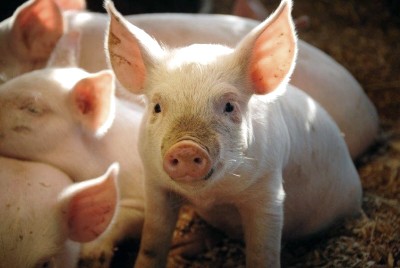
Respiratory research faces a persistent translation problem. Molecules and devices that perform well in rodents frequently fail when they reach human trials, largely because the rodent lung differs from ours in size, branching pattern, and immune behavior. The pig closes much of that gap. With lungs that resemble human organs in anatomy and physiology, pigs allow researchers to run bronchoscopy, mechanical ventilation, and advanced imaging in a living system that behaves far more like a patient than a cage of mice ever could. Drawing on more than two decades of hands-on large animal model experience at Biotech Farm, this article explains what pig model respiratory research involves, how porcine lung and airway models are built, which readouts they generate, and how teams plan and validate these studies for maximal translational value.
Exclusive Insight: The single most common reason porcine respiratory studies underdeliver is not the model — it is protocol drift. Teams that lock endpoints, randomization, and SOPs before the first animal is enrolled consistently produce data that survives regulatory scrutiny; teams that improvise mid-study rarely do.
Why the Pig Has Become a Cornerstone of Respiratory Research
Pig model respiratory research is the use of pigs, whether living, organ-level, or tissue-level preparations, to study the physiology and pathology of the respiratory system. The approach spans infectious disease, acute lung injury, airway hyperresponsiveness, cystic fibrosis, and the evaluation of inhaled or implanted therapies. The rationale is straightforward: the porcine lung shares with the human lung a similar tracheobronchial tree, comparable alveolar architecture, and pulmonary function values that scale predictably to human ranges. A recent overview of swine models in respiratory infections concluded that pigs offer substantial value across pathogens and conditions, while also noting that protocol harmonization remains an open need in the field — a point any serious study sponsor should internalize early.
Pig airways branch in a pattern closer to the human dichotomous scheme than to the monopodial branching of rodents, which changes airflow, particle deposition, and drug distribution in ways that matter for inhaled therapies. Lung size permits use of clinical-grade catheters, endotracheal tubes, ventilators, and imaging hardware. The immune system of the pig also responds to respiratory pathogens with cytokine profiles that track human responses reasonably well — the same foundation that supports the classic argument that the porcine lung can serve as a model for cystic fibrosis lung disease.
What Is a Pig Airway Model and What Does It Measure?
A pig airway model narrows the focus to the trachea, bronchi, and bronchioles. Researchers use it to study airflow dynamics, airway reactivity, inflammation, infection, and biofilm formation — the core processes behind asthma, COPD, cystic fibrosis, and respiratory infections. Because porcine airway epithelium closely resembles human epithelium, these models support detailed analysis of mucus production, ciliary beat function, and epithelial responses to challenge. Ex vivo airway tissue preparations can quantify microbial growth, virulence expression, and tissue damage in chronic disease settings, which is exactly the kind of data that cell cultures cannot provide on their own.
What Is a Porcine Lung Model in Practice?
A porcine lung model is any experimental system built on intact or partial pig lung tissue to study respiratory, infectious, or inflammatory responses under controlled conditions. The family includes in vivo studies in living animals, ex vivo perfused and ventilated lungs, and precision-cut lung slices maintained in culture. Each tier answers a different question. Disease mechanism work, therapeutic testing, and medical device evaluation all draw on these formats, and the choice among them is one of the most consequential decisions a study team makes.
In Vivo, Ex Vivo, or In Vitro: Which Format Fits Your Question?
The three formats differ in physiological relevance, control, and cost, and the trade-offs deserve honest assessment rather than default habit.
| Format | What It Is | Main Strengths | Main Limitations |
|---|---|---|---|
| In vivo | Whole living pig under anesthesia or awake instrumentation | Systemic immune responses, long-term outcomes, full physiology | Higher cost, ethical burden, less isolation of variables |
| Ex vivo | Isolated lung on perfusion and ventilation, such as EVLP | Tight control of perfusate and ventilation, detailed sampling, fewer animals | Limited duration, no systemic immunity |
| In vitro | Cell cultures or precision-cut lung slices from pig lung | High throughput, low cost, precise microenvironment control | No tissue architecture at organ scale, no systemic effects |
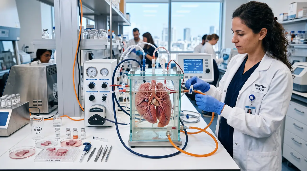
Ex vivo models earn their place when the research question is localized to the lung itself. Drug screening, pharmacokinetics of locally delivered agents, and mechanism-of-action studies all benefit from the ability to set perfusion pressure, ventilation parameters, and drug concentration exactly, then sample the tissue directly. Variability tends to drop because the systemic compartment, with its hormonal and neural influences, is removed from the equation. Teams exploring the broader landscape can find a wider survey of applications on the swine biomedical models resource page, which covers formats beyond the respiratory arena.
When Is a Living Pig the Only Acceptable Option?
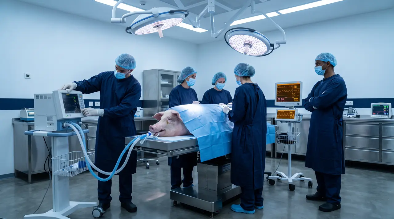
Some questions cannot be answered without an intact organism. Systemic immune responses to infection or vaccination, long-term safety of implanted devices, survival endpoints, and interactions between the lung and other organ systems all demand in vivo work. A therapy that stabilizes an isolated lung may still fail because of cardiovascular side effects, hemodynamic instability, or systemic inflammation, and only a whole animal reveals that. In practice, many respiratory device and drug programs pair in vivo pulmonary work with parallel whole-animal cardiovascular evaluation, similar in logic to what is described for pig model cardiovascular device testing, because the systems interact continuously in a living patient.
Swine Respiratory Disease Models: Pathogens and Conditions
A swine respiratory disease model simulates or induces respiratory disease in pigs to study pathology, diagnosis, or treatment efficacy. Its users range from pharmaceutical and biotech companies to academic groups and medical device developers. Bacterial models include Mycoplasma hyopneumoniae for chronic respiratory disease, Actinobacillus pleuropneumoniae for acute pleuropneumonia, Pasteurella multocida as a secondary invader, and Bordetella bronchiseptica in rhinitis. On the viral side, influenza A in swine is highly relevant to human pandemic preparedness, while porcine reproductive and respiratory syndrome virus and porcine circovirus type 2 round out the major veterinary pathogens with research value. Non-infectious conditions modeled in pigs include acute respiratory distress syndrome, cystic fibrosis through genetically modified lines, airway hyperresponsiveness, and pulmonary fibrosis.
The overlap with human-relevant pathology is what turns veterinary knowledge into translational opportunity — pigs offer substantial value across respiratory pathogens and conditions, but protocol harmonization remains an open need in the field.
— Overview of swine models in respiratory infection research
Which Readouts Can You Actually Collect From Pig Respiratory Studies?
The readout menu in pigs is broad because the animal is large enough for clinical instruments. Physiological measurements include lung compliance and resistance, blood gas analysis such as the PaO2/FiO2 ratio, oxygen saturation, and full ventilation parameters. Imaging spans radiography, CT, and ultrasound for structural and functional assessment. Inflammatory profiling covers cytokines and chemokines in bronchoalveolar lavage fluid, tissue homogenates, and blood. Microbiological work includes bacterial load in colony-forming units, viral titers, pathogen identification, and microbiome analysis. Histopathology rounds out the package with scoring of injury, inflammation, fibrosis, and remodeling.
Physiological
- Lung compliance and resistance
- PaO2/FiO2 ratio and blood gases
- Ventilation mechanics: PIP, plateau, compliance
Immunological
- BAL fluid cell counts and cytokines
- Tissue homogenate inflammatory markers
- Systemic serum profiling
Structural & Microbial
- CT, radiography, and ultrasound imaging
- Bacterial load in CFU and viral titers
- Standardized histopathology scoring
Whole Lung or Precision-Cut Lung Slices: How to Decide?
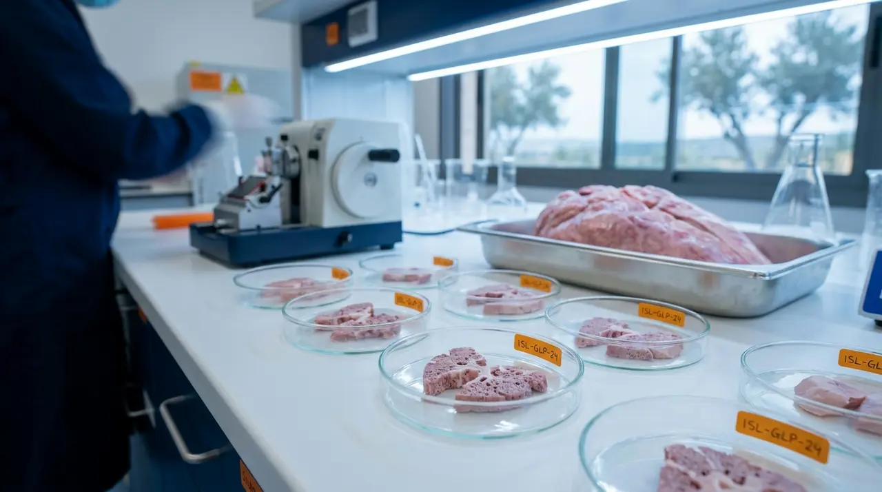
Precision-cut lung slices, or PCLS, are thin slices of viable lung tissue maintained in culture, typically for days to weeks. They excel at rapid screening, high-throughput drug testing, and mechanistic studies of cellular responses. Studies in porcine PCLS have demonstrated functional readouts such as airway contraction in response to methacholine, which shows that even tissue-level preparations retain relevant physiology. Their limitation is equally clear: no perfusion, no ventilation at organ scale, no immune system, no systemic effects. If the research question involves ventilation-induced injury, hemodynamics, or whole-body responses, only whole-lung formats, in vivo or ex vivo, will answer it. The decision should follow the question, not the available equipment.
What Is Ex Vivo Lung Perfusion in a Pig Model?
Ex vivo lung perfusion, or EVLP, is a technique in which an isolated pig lung is perfused with a physiological solution and ventilated outside the body, maintaining viability and function for hours. The core setup includes a perfusion circuit, a mechanical ventilator, and temperature control, together creating a stable platform for intervention. Applications include assessment of lung viability in the transplantation context, study of drug delivery and absorption directly into lung tissue, evaluation of lung protection strategies, and investigation of acute lung injury under tightly controlled conditions. Physiological reviews of EVLP across species highlight the pig as a particularly useful preclinical platform because organ size and vascular anatomy match clinical hardware.
How Do You Plan a Pig Pulmonary Study That Does Not Fail?
Study planning is where translational programs are won or lost. The essentials are unglamorous but decisive: defined primary and secondary endpoints chosen before the first animal is enrolled, a model format matched to the research question, statistical power analysis with honest sample size estimates, pilot experiments to validate feasibility, and standard operating procedures for every procedure from intubation to tissue fixation. Complex models such as ARDS and respiratory infection make these disciplines non-negotiable, because without predefined clinical and laboratory parameters, reproducibility and interpretation both collapse.
| Planning Item | Why It Matters |
|---|---|
| Ethical and regulatory approval | Committee review is mandatory and shapes what the protocol may contain |
| Animal source and health status | Undocumented pathogen exposure can silently corrupt infection studies |
| Randomization and blinding | Bias control, especially in subjective endpoints like histology scoring |
| Data management plan | Predefined collection and storage prevents post hoc cherry-picking |
| Experienced team | Large animal handling, veterinary care, and respiratory physiology skills are distinct crafts |
Methodology: How Study Design Decisions Are Made at an Experienced Facility
At an established large animal facility, model selection begins with the endpoint, not the animal. The team works backward from the translational claim the sponsor needs to make: if the claim concerns systemic safety, an in vivo design is drafted; if it concerns local drug deposition or lung-protective ventilation strategies, an ex vivo EVLP circuit is specified. Protocol development proceeds through feasibility review with veterinary staff, ethical submission support, and pilot runs that validate intubation, ventilation, and sampling procedures before the pivotal study begins.
This staged methodology — endpoint first, model second, protocol third — is what separates data that survives regulatory scrutiny from data that merely fills a report. In Israel specifically, sourcing pigs for research involves documented health declarations and regulated welfare conditions, which is one reason working with an established facility simplifies the early phase of a project considerably.
How Do Researchers Validate a Porcine Respiratory Model?
Validation is the process of demonstrating that the model faithfully reflects the human condition it is meant to represent. It has several layers. Phenotypic similarity means the model shows clinical signs, pathological changes, and physiological dysfunctions comparable to the human disease. Molecular similarity requires that gene expression patterns, immunological responses, and biomarker profiles mirror those in patients. Response to standard therapies is a particularly persuasive test: if a treatment with known human efficacy works in the pig and one with known failure does not, the model earns credibility. Reproducibility across experiments and sites completes the picture.
Case Example: From Model Development to Prevention Research
Recent work illustrates the translational trajectory well. Researchers developing new pig models for respiratory disease prevention created a framework for testing immunoprophylactic interventions aimed at reducing pathogen transmission, work reported as an advance in pig model development for respiratory disease prevention.
The pattern is a familiar one in the field: a model is established and characterized, validated against known disease parameters, and then applied to candidate interventions whose human relevance depends entirely on the quality of that earlier validation. Teams that respect the sequence get answers they can trust.
Where Pig Models Fall Short — Know Before You Commit: Costs and logistics run higher than in rodents, from housing to anesthesia time. Genetic diversity adds variability, though it can also be defended as more representative of human populations. Ethical scrutiny is more intense given the size and cognitive sophistication of pigs. And pigs are not humans: differences in drug metabolism and fine details of immune response exist and must be checked for each compound. Acknowledging these limits in a regulatory submission strengthens rather than weakens the program.
What Are the Ethical Obligations in This Field?
Ethical practice in pig pulmonary research rests on the 3Rs: replacement with non-animal methods where possible, reduction of animal numbers through sound statistics and shared controls, and refinement of procedures to minimize pain and distress. Animal welfare standards cover housing, husbandry, anesthesia, analgesia, and humane endpoints. Every protocol must justify that the scientific benefit outweighs the ethical cost, and that justification must be substantive, not formulaic. Facilities that document welfare practices transparently also protect their research partners, because ethical gaps discovered late can invalidate years of data.
Why Does the Choice of Research Facility Matter So Much?
Large animal respiratory research is operationally demanding in ways that small animal work is not. It requires surgical suites sized and equipped for pigs, ventilators and imaging capable of clinical-grade measurement, veterinarians experienced in porcine medicine, and staff who can handle animals safely and humanely. A facility like Biotech Farm brings together surgical infrastructure, imaging tools such as fluoroscopy and high-definition ultrasound, and a professional crew with decades of experience in leading large animal studies. For teams without in-house large animal capability, that combination converts a project from theoretically possible to practically executable.
The practical value of a specialist facility lies in the details: matching study design to the available models, help with protocol development before ethical submission, veterinary oversight during procedures, and structured data collection that survives later scrutiny. Cost components include animal acquisition and husbandry, surgical and veterinary care, specialized equipment and consumables, personnel time, and data analysis and reporting. An ex vivo perfusion study of a few hours differs enormously from a twelve-week in vivo device evaluation, so budgets and schedules must be built per design, not from a price list.
Practical Tip: Early consultation with an experienced facility is usually the fastest way to get a realistic budget and timeline picture before committing resources — and it frequently prevents methodological flaws that would otherwise surface only at the analysis stage.
Frequently Asked Questions
Where Should Your Next Respiratory Study Begin?
So the question worth asking is this: which respiratory research question in your pipeline would benefit most from a model that actually behaves like a human lung? Whether you are evaluating an inhaled therapy, testing a ventilation strategy, characterizing an infection model, or developing a device that interfaces with the airway, the right porcine model can move you closer to the clinic with far less translational risk.